
Objective: The primary objective was to determine whether early pre-synaptic caudate denervation, measured using 123 I ioflupane SPECT quantitative metrics, provides independent and incremental prognostic value for future cognitive decline in Parkinson’s disease (PD) beyond usual established clinical risk factors.
Background: Cognitive impairment affects patients with PD and progresses heterogeneously. Given the key role of the caudate nucleus in the fronto-striatal cognitive circuitry, early pre-synaptic caudate denervation, measured using dopamine transporter (DaT) quantitative metrics, may provide a surrogate measure of future cognitive impairment.
Method: We performed a longitudinal analysis of participants with PD in the Parkinson’s Progression Markers Initiative (PPMI). The primary outcome was development of Mild Cognitive impairment (MCI) on follow-up. A hierarchical Cox proportional hazards model was applied, beginning with an anchor clinical model. Regional DaT scan metrics from predefined striatal subregions were sequentially added to assess incremental prognostic utility.
Results: Among 676 cognitively normal participants followed up for a median of 3.0 years, 178 (26.3%) developed incident MCI. In the multivariable clinical anchor model (C-index=0.647), older age of onset (HR 1.03, p=0.0007) and higher autonomic dysfunction (SCOPA-AUT; HR 1.04, p=0.004) were independent predictors of MCI, while higher education was protective (HR 0.94, p=0.044). Disease duration and motor severity (Hoehn–Yahr/PIGD) were not significant. Adding DaTSCAN measures (N=646) improved discrimination, with higher ipsilateral caudate uptake significantly reducing MCI risk (HR 0.81 per SD, 95% CI 0.67–0.98; p=0.032; LRT p=0.031). Higher mean caudate uptake was similarly predictive (HR 0.83 per SD, 95% CI 0.70–1.00; p=0.046), whereas putaminal binding and other striatal measures were not significantly associated with incident MCI.
Conclusion: Caudate dopaminergic denervation at baseline independently predicts incident MCI in PD and assists early cognitive risk stratification.
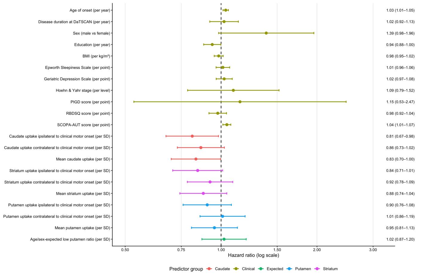
Forest plot of clinical and imaging predictors
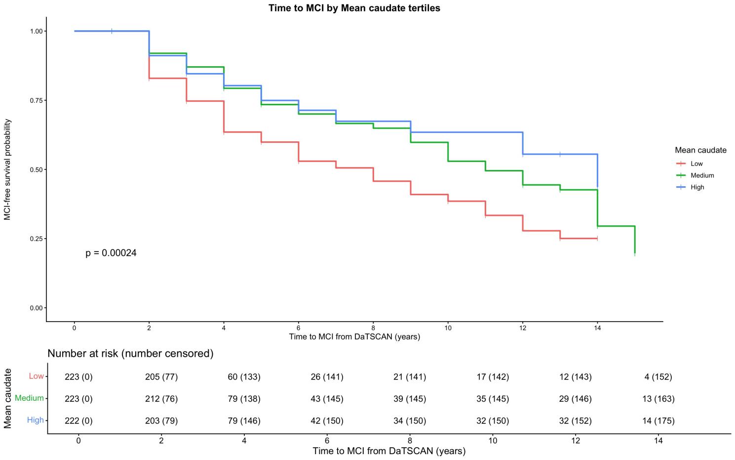
K-M curves stratified by mean caudate DaT uptake
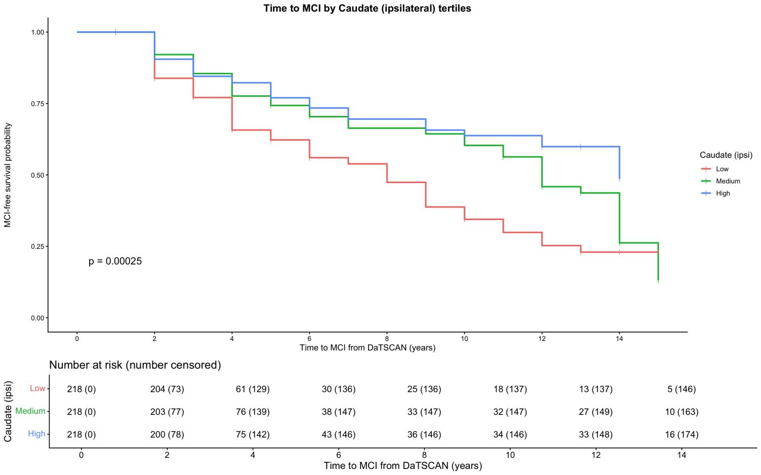
K-M curves stratified by ipsilateral caudate DaT
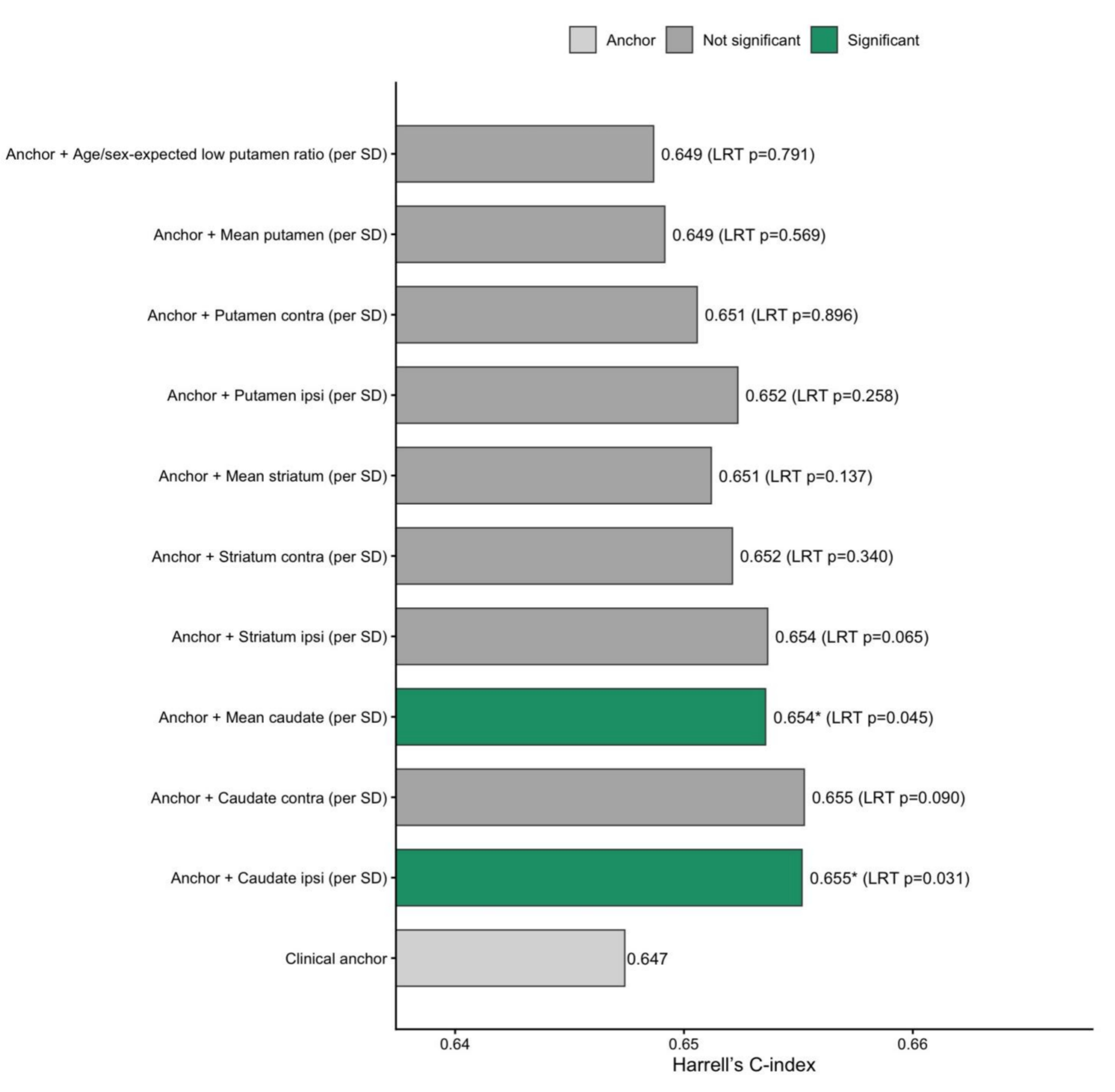
Incremental prognostic value of DAT imaging
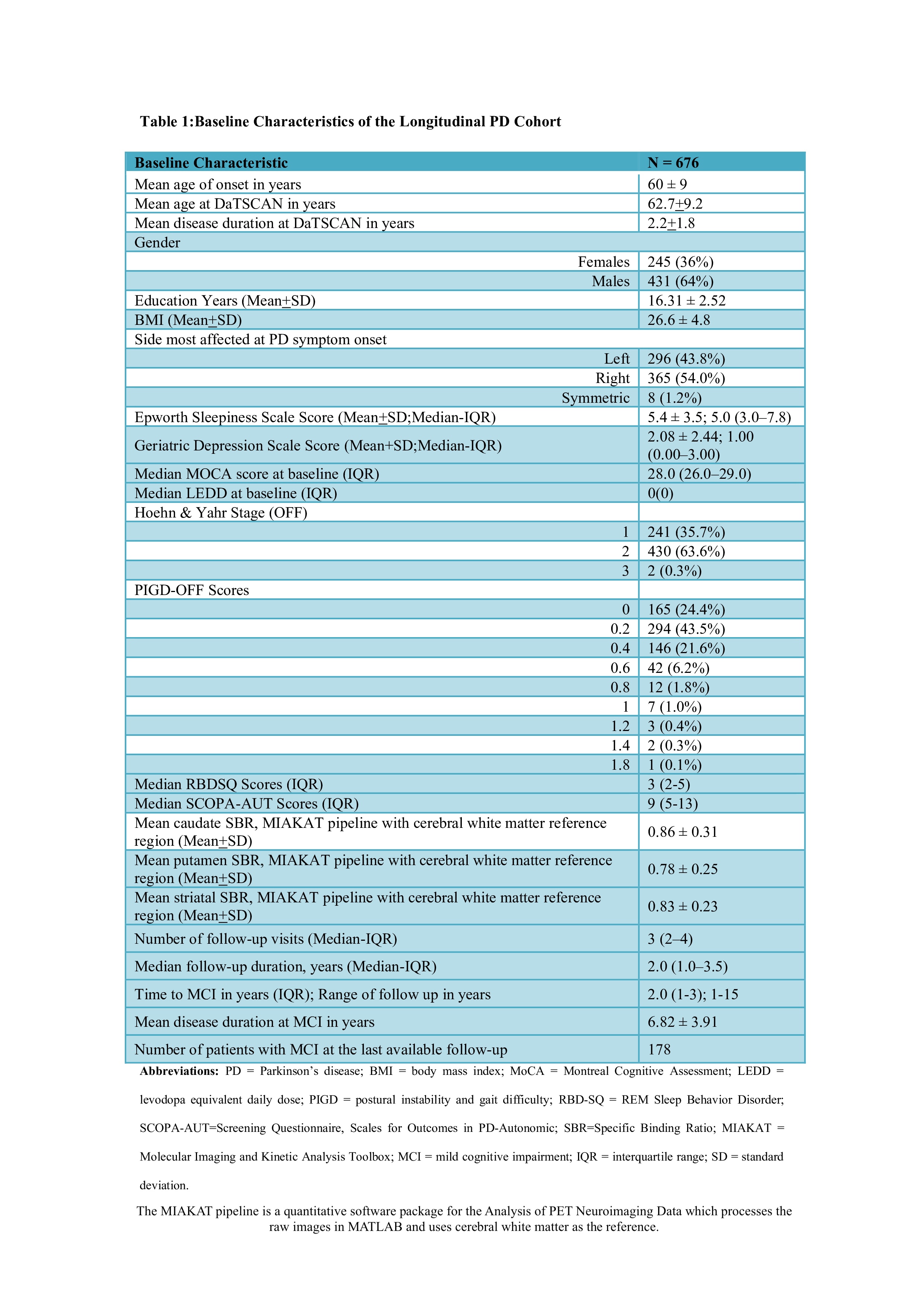
Table:Baseline Characteristics
References: 1. Pasquini J, Durcan R, Wiblin L, et al. Clinical implications of early caudate dysfunction in Parkinson’s disease. Journal of Neurology, Neurosurgery & Psychiatry 2019;90:1098-1104.
2. Ramezani, F., Mahmoudi, S., & Safari, F. (2025). Association between DATSCAN-derived dopaminergic activity and cognitive performance and quality of life in Parkinson’s disease: A cross-sectional study. eNeurologicalSci, 100599. https://doi.org/10.1016/j.ensci.2025.100599
To cite this abstract in AMA style:
B. Panigrahi, D. Radhakrishnan, R. Rajan. Early Caudate Dopaminergic Loss as an Imaging Biomarker of Cognitive Impairment in Parkinson’s Disease: A Longitudinal Analysis [abstract]. Mov Disord. 2026; 41 (suppl 1). https://www.mdsabstracts.org/abstract/early-caudate-dopaminergic-loss-as-an-imaging-biomarker-of-cognitive-impairment-in-parkinsons-disease-a-longitudinal-analysis/. Accessed October 1, 2026.« Back to 2026 International Congress
MDS Abstracts - https://www.mdsabstracts.org/abstract/early-caudate-dopaminergic-loss-as-an-imaging-biomarker-of-cognitive-impairment-in-parkinsons-disease-a-longitudinal-analysis/