
Objective: To investigate gray matter (GM) volume changes in sporadic dystonia (SD) using T1 MRI and voxel-based morphometry (VBM).
Background: Dystonia is a hyperkinetic movement disorder with sustained or intermittent muscle contractions producing abnormal, repetitive movements and postures. Neuroimaging studies have identified metabolic and functional network abnormalities within basal ganglia-cerebellar-cortical motor networks in hereditary dystonia [1]. However, the structural brain changes underlying sporadic forms remain incompletely characterized.
Method: Twenty patients with SD and 29 HC were recruited through Center for Neurosciences, Northwell Health. Diagnosis of SD was confirmed using established criteria. T1 MRI was acquired on a Siemens 3T Prisma using 3D MPRAGE. GM volume differences were assessed using FSL-VBM. [2-6].Voxel-wise group comparisons were conducted using permutation testing. Significant clusters were labeled with the AAL atlas [7], and GM volumes were extracted for post-hoc analysis. Motor symptoms severity was evaluated by a neurologist in the off-medication state at the time of imaging using the Burke–Fahn–Marsden Dystonia Rating Scale (BFMDRS).
Results: Demographic analysis revealed no significant differences between groups in age and gender (p=0.49 and 0.64, respectively) [Table 1].VBM analysis revealed that SD showed significantly reduced GM volume compared to HC in three clusters [Table 2; Fig 1]. The largest encompassed the right lingual gyrus, right cerebellum, vermis, bilateral thalamus, and posterior cingulate cortex. Post-hoc analysis revealed significant reductions in the right fusiform gyrus, right cerebellum, left thalamus and left posterior cingulate (p<0.05) [Fig 2]. Exploratory correlation analyses revealed no significant associations between GM volumes and BFMDRS motor scores (6.3±3.7) and disability score (1.7±2.5) (p>0.05).
Conclusion: Patients with SD demonstrated significant GM volume reductions in regions that are consistent with prior evidence in the dystonia pathophysiology and further suggest that structural abnormalities in visual and higher order associative cortices may also contribute to the disorder. By elucidating the neuroanatomical substrates associated with SD, these regions may serve as potential imaging biomarkers and help guide therapeutic interventions.
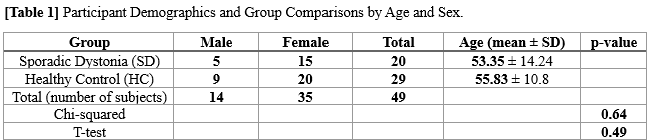
Demographics and Group Comparisons by Age and Sex
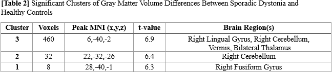
Clusters: Significant GM Volume Differences
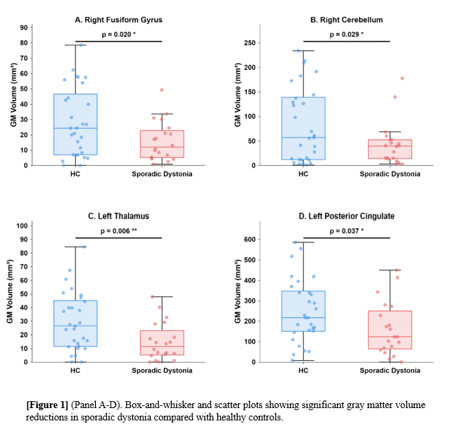
Significant GM volume reductions between groups
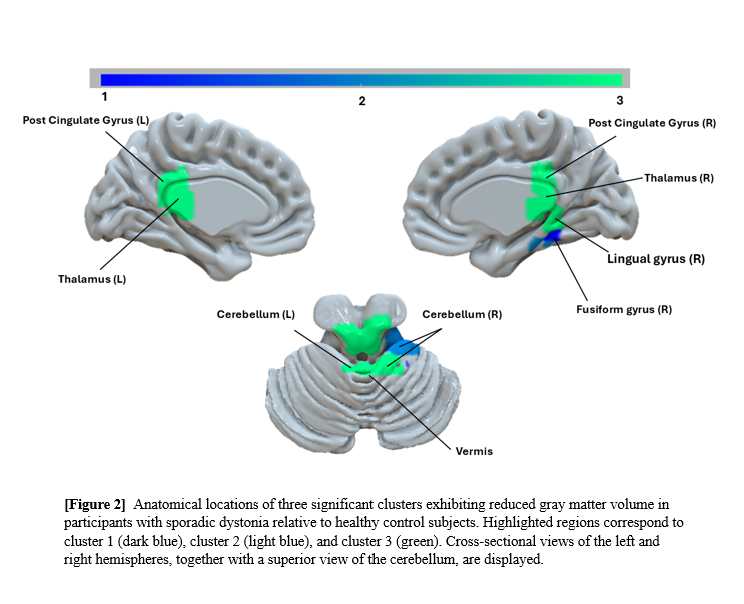
Anatomical locations of cluster brain regions
References: 1. An Vo, Nha Nguyen, Koji Fujita, Katharina A Schindlbeck, Andrea Rommal, Susan B Bressman, Martin Niethammer, David Eidelberg, Disordered network structure and function in dystonia: pathological connectivity vs. adaptive responses, Cerebral Cortex, Volume 33, Issue 11, 1 June 2023, Pages 6943–6958,
2. Rajagopalan V, Pioro EP. Disparate voxel based morphometry (VBM) results between SPM and FSL softwares in ALS patients with frontotemporal dementia: which VBM results to consider? BMC Neurol. 2015 Mar 13;15:32. doi: 10.1186/s12883-015-0274-8. PMID: 25879588; PMCID: PMC4371611.
3. Douaud G, Smith S, Jenkinson M, Behrens T, Johansen-Berg H, Vickers J, James S, Voets N, Watkins K, Matthews PM, James A (2007) Anatomically related grey and white matter abnormalities in adolescent-onset schizophrenia. Brain 130:2375-2386
4. Good CD, Johnsrude IS, Ashburner J, Henson RN, Friston KJ, Frackowiak RS (2001) A voxel-based morphometric study of ageing in 465 normal adult human brains. NeuroImage 14:21-36.
5. Smith SM, Jenkinson M, Woolrich MW, Beckmann CF, Behrens TE, Johansen-Berg H, Bannister PR, De Luca M, Drobnjak I, Flitney DE, Niazy RK, Saunders J, Vickers J, Zhang Y, De Stefano N, Brady JM, Matthews PM (2004) Advances in functional and structural MR image analysis and implementation as FSL. NeuroImage 23 Suppl 1:S208-219.
6. Andersson, M. Jenkinson and S. Smith (2007) Non-linear registration, aka Spatial normalisation. FMRIB technical report TR07JA2
7. Tzourio-Mazoyer, N., Landeau, B., Papathanassiou, D., Crivello, F., Etard, O., Delcroix, N., & Mazoyer, B. (2002). Automated anatomical labeling of activations in SPM using a macroscopic anatomical parcellation of the MNI MRI single-subject brain. NeuroImage, 15(1), 273-289.
Acknowledgments: This work was funded by the Dystonia Medical Research Foundation (DMRF)
and the Career Enhancement Award (CEA) to An Vo. We are grateful to all study
participants and research staff who contributed to data collection.
To cite this abstract in AMA style:
O. Omilabu, S. Vacca, T. Fitzpatrick, M. Neithammer, N. Nguyen, D. Eidelberg, A. Vo. Voxel-Based Morphometry Reveals Gray Matter Volume Reductions in Sporadic Dystonia [abstract]. Mov Disord. 2026; 41 (suppl 1). https://www.mdsabstracts.org/abstract/voxel-based-morphometry-reveals-gray-matter-volume-reductions-in-sporadic-dystonia/. Accessed October 1, 2026.« Back to 2026 International Congress
MDS Abstracts - https://www.mdsabstracts.org/abstract/voxel-based-morphometry-reveals-gray-matter-volume-reductions-in-sporadic-dystonia/