
Category: Parkinson's disease: Neuroimaging
Objective: This study aimed to identify the alterations of local synchronization and functional connectivity in PD and explore the acute neural response to acupuncture stimulation using resting-state fMRI.
Background: Parkinson’s disease (PD) is a neurodegenerative disease characterized by a broad spectrum of motor and non-motor symptoms arising from the disruption of complex neural networks. Identifying functional alterations has become increasingly essential for a more comprehensive understanding of its underlying mechanisms. The integration of Regional homogeneity (ReHo) and functional connectivity (FC) analyses can provide a comprehensive insight for elucidating the underlying neural substrates of Parkinson’s disease.
Method: This study used resting-state fMRI to identify the functional alterations between 48 patients with PD and 24 healthy controls. All participants with PD were evaluated for the severity of symptoms using the Unified Parkinson’s Disease Rating Scale and Balance Master. Regional Homogeneity (ReHo) was calculated by measuring the temporal synchronization between the time series of a given voxel and those of its nearest neighbors (27 voxels) using Kendall’s Coefficient of Concordance on a voxel-wise basis. Based on the significant group differences in ReHo maps, seed-based functional connectivity analysis (z-transformed) was conducted, identifying further distinct connectivity alterations between the two groups. Furthermore, the performance of the identified zFC in classifying PD and healthy controls was evaluated through a binomial logistic regression.
Results: The PD group exhibited increased ReHo values in the left middle frontal gyrus. Furthermore, seed-based zFC analysis, using this region as a seed, revealed decreased connectivity with the left inferior parietal region in the PD group. In the binomial logistic regression applying zFC values of identified regions, age, and sex, as covariates, the statistics were as follows: accuracy 0.90, specificity 0.88, sensitivity 0.92, and AUC 0.95.
Conclusion: This study identified the altered local synchronization in the left middle frontal gyrus of patients with Parkinson’s disease and demonstrated that the functional connectivity between the left middle frontal and left inferior parietal regions may serve as a key neural substrate for distinguishing PD from healthy controls.
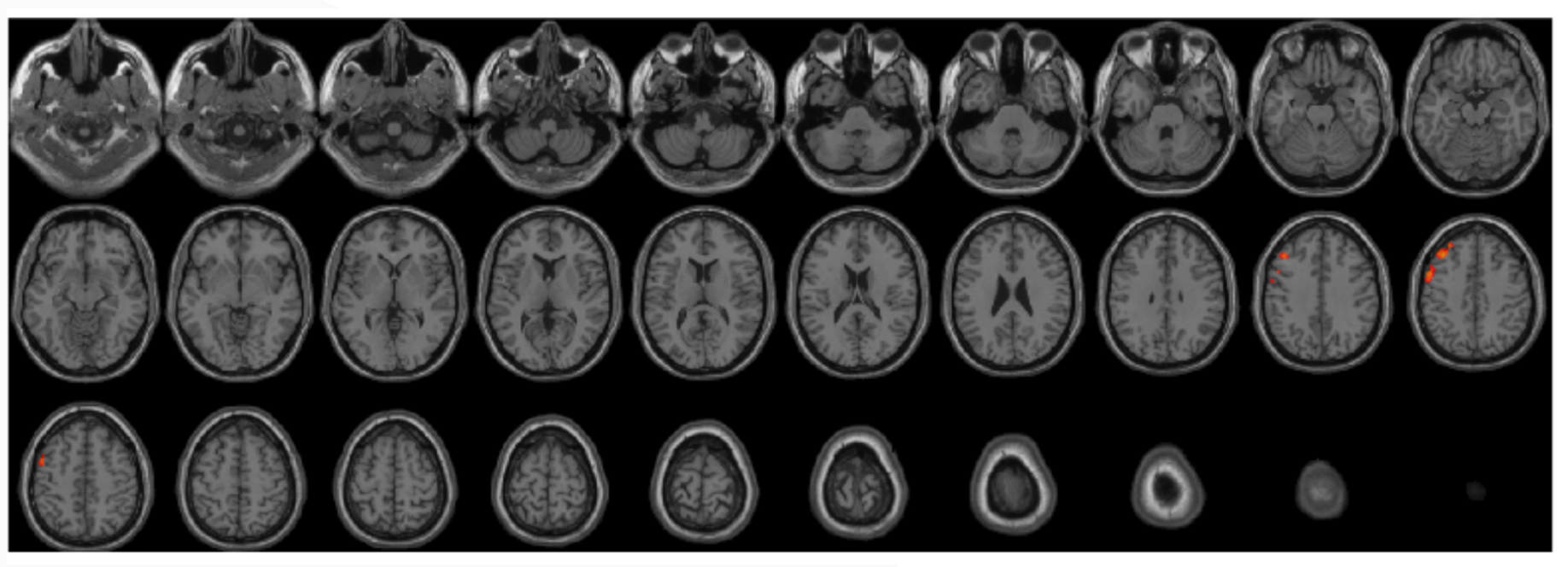
Results of regional homogeneity between PD and HC
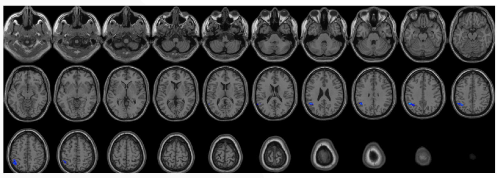
Seed based zFC of middle frontal gyrus
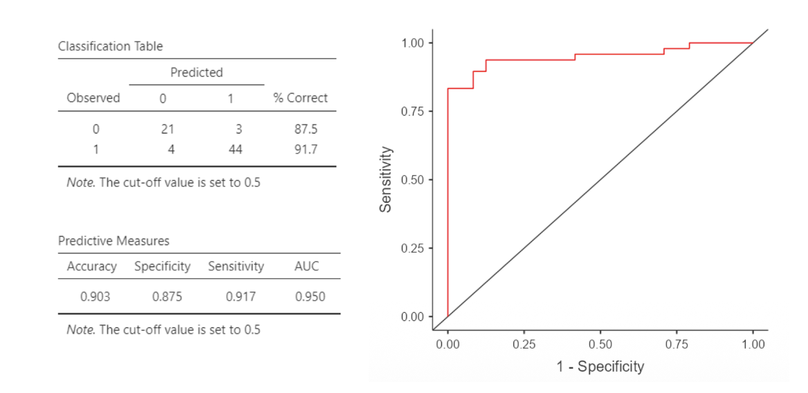
Binomial logistic regression model
To cite this abstract in AMA style:
DH. Lee. Altered local synchronization and functional connectivity in Patients with Parkinson’s Disease [abstract]. Mov Disord. 2026; 41 (suppl 1). https://www.mdsabstracts.org/abstract/altered-local-synchronization-and-functional-connectivity-in-patients-with-parkinsons-disease/. Accessed October 1, 2026.« Back to 2026 International Congress
MDS Abstracts - https://www.mdsabstracts.org/abstract/altered-local-synchronization-and-functional-connectivity-in-patients-with-parkinsons-disease/