
Objective: The present study aimed to quantitatively compare reflexive and voluntary saccades in patients with Parkinson’s disease (PD) and multiple system atrophy (MSA), and to examine task-dependent modulation of saccadic parameters across disorders.
Background: Saccadic eye movements are classified into reflexive and voluntary paradigms, controlled by distinct neural circuits. Reflexive saccades are mediated by parietal-superior colliculus-brainstem pathways, whereas voluntary saccades require internally guided motor control involving frontal cortical networks. Previous studies have shown that PD has preserved reflexive saccades, but impaired voluntary saccades with prolonged latency and hypometric amplitude. Given that MSA involves degeneration including cerebellar and brainstem structures, their saccadic movements may differ from those in PD.
Method: We prospectively enrolled 22 PD patients (65.09 ± 7.09 years), 13 MSA patients (67.15 ± 7.21 years) and healthy controls (63.44 ± 7.98 years). Participants underwent video-oculography assessing reflexive and voluntary saccades. Saccadic gain, peak velocity and latency were analyzed for reflexive saccades, whereas gain, peak velocity and intersaccadic pause in the voluntary paradigm.
Results: Reflexive saccades showed preserved gain and peak velocity across groups, although latency was significantly prolonged in PD and MSA. Voluntary saccades revealed more pronounced abnormalities in both patient groups, including hypometric gain, reduced peak velocity and prolonged intersaccadic pause. A significant Group x Paradigm interaction was observed for saccadic gain. Healthy controls showed increased gain in voluntary paradigm, while PD exhibited attenuated modulation and MSA showed markedly reduced or reversed modulation. A significant interaction was also observed for peak velocity. The Group x Paradigm interaction remained significant after adjustment for age.
Conclusion: Voluntary saccadic paradigms reveal more prominent abnormalities than reflexive saccades in parkinsonian disorders. Task-dependent modulation of saccadic gain, in particular, differentiated controls from PD and MSA, and may reflect disruption of cerebellar and frontal-basal ganglia circuits involved in internally guided eye movements. These findings suggest that voluntary saccade testing may provide mechanistic insight into differential network involvement in parkinsonian disorders.
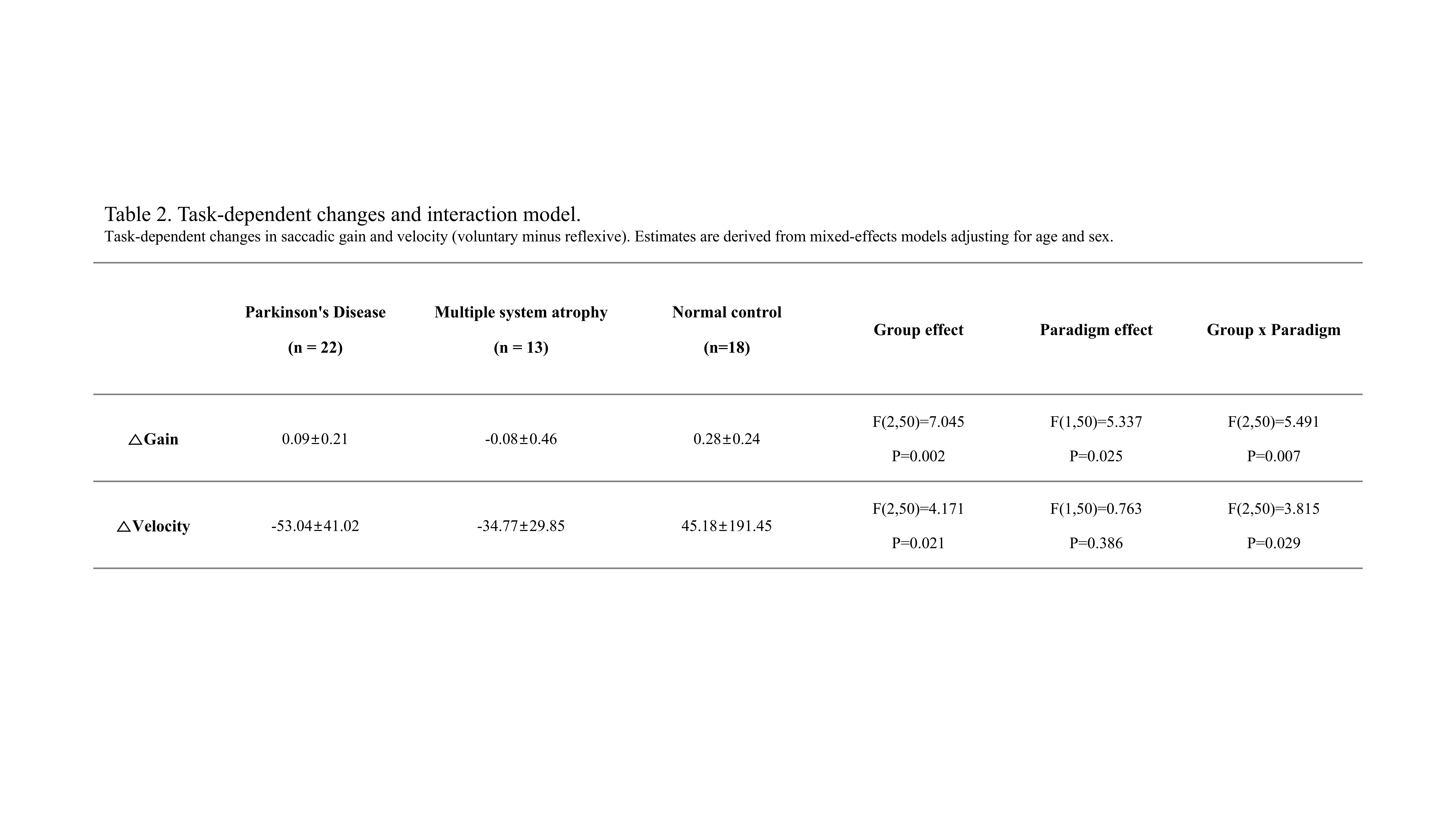
Table 1.
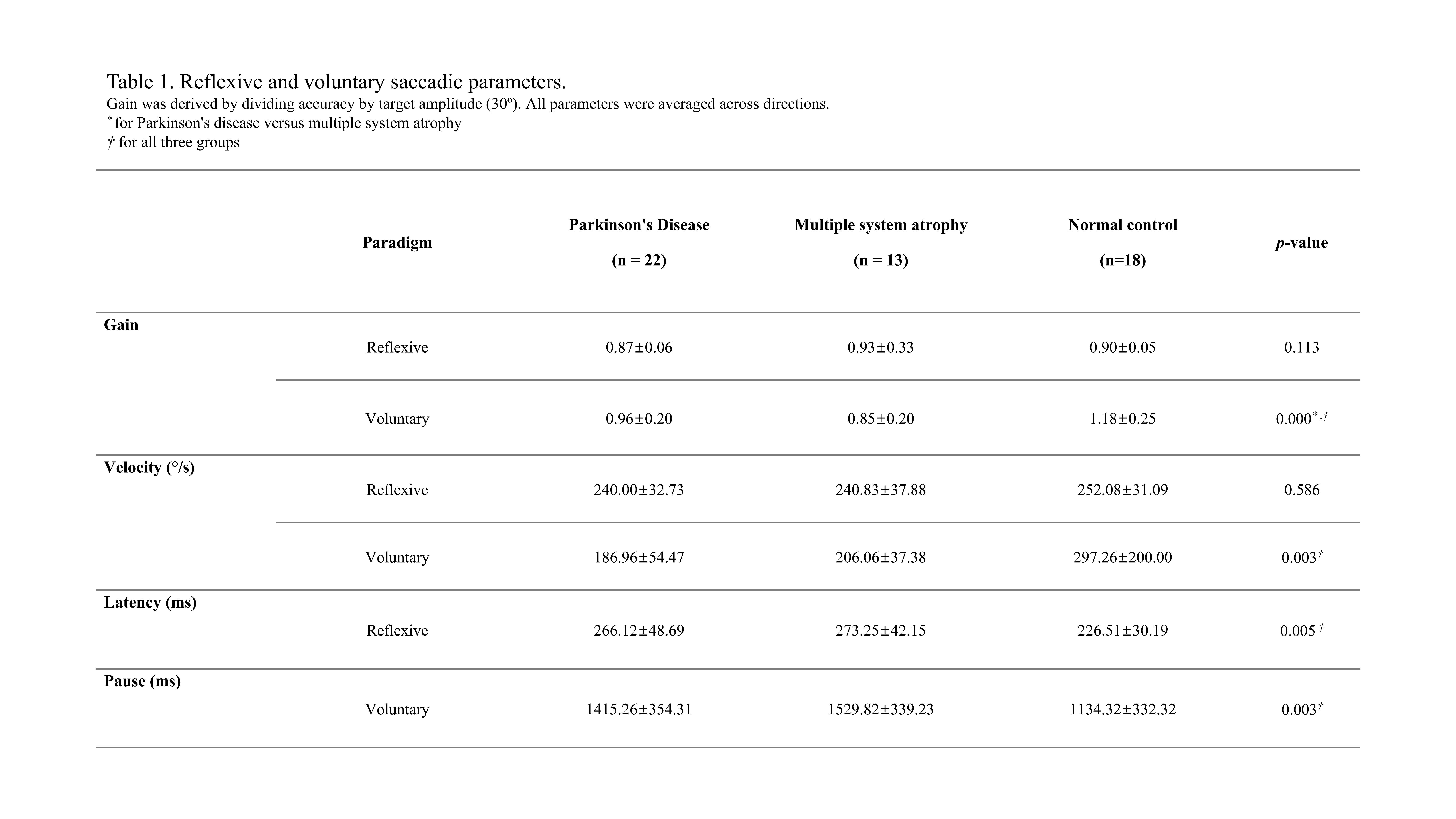
Table 2.
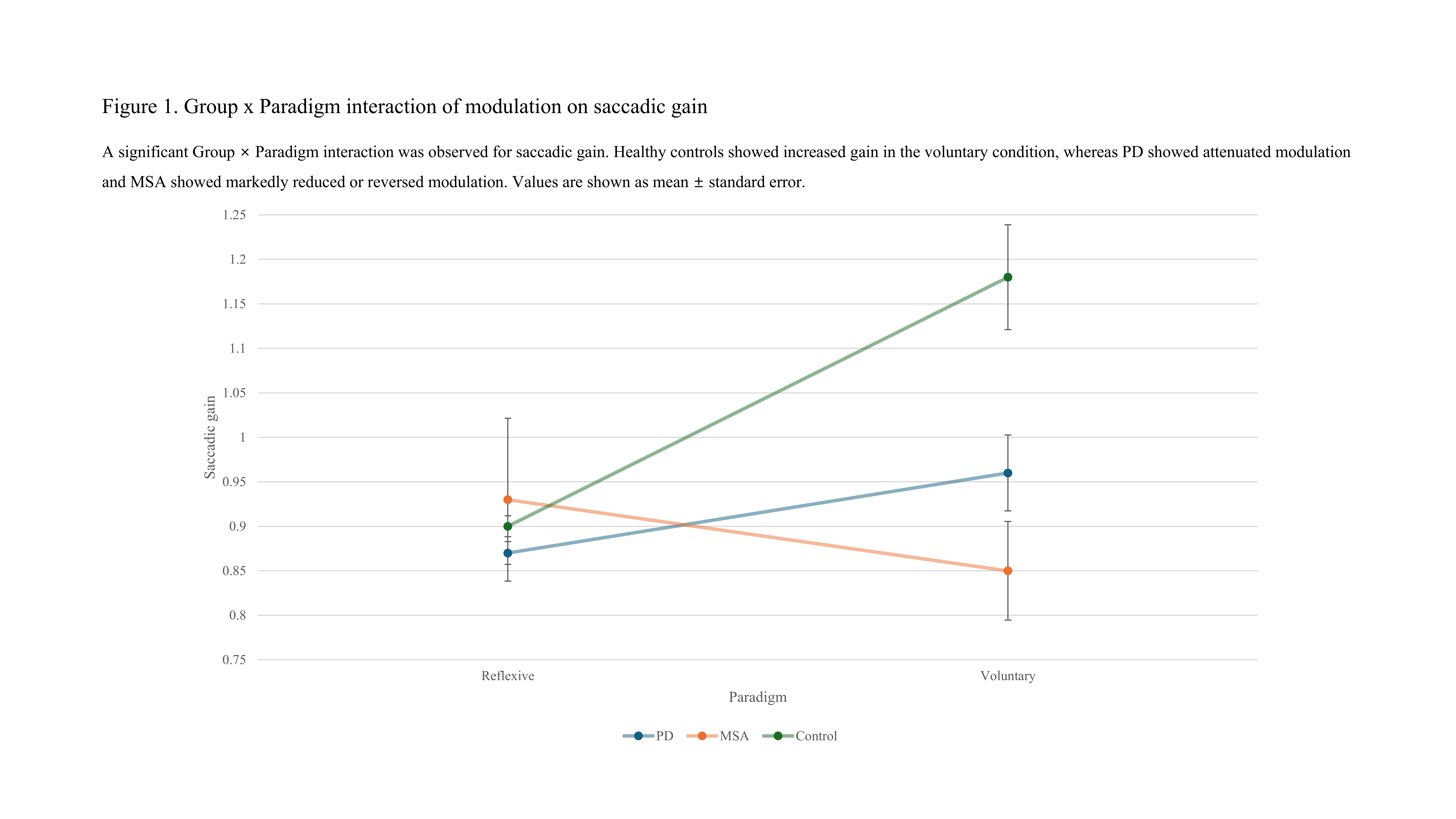
Figure 1.
To cite this abstract in AMA style:
H. Kim, Y. Bae, J. Yun. Task-Dependent Modulation of Reflexive and Voluntary Saccades in Parkinson Disease and Multiple System Atrophy [abstract]. Mov Disord. 2026; 41 (suppl 1). https://www.mdsabstracts.org/abstract/task-dependent-modulation-of-reflexive-and-voluntary-saccades-in-parkinson-disease-and-multiple-system-atrophy/. Accessed October 1, 2026.« Back to 2026 International Congress
MDS Abstracts - https://www.mdsabstracts.org/abstract/task-dependent-modulation-of-reflexive-and-voluntary-saccades-in-parkinson-disease-and-multiple-system-atrophy/