
Category: Rare Neurometabolic Movement Disorders
Objective: We used cellular and mouse models to explore the subcellular origin of calcification foci caused by primary brain calcification (PBC) gene defects (SLC20A2 and XPR1).
Background: PBC is a neurological disorder characterized by symmetric bilateral calcification in the basal ganglia regions, exhibiting high genetic and clinical heterogeneity. Identification of multiple pathogenic genes has advanced pathogenesis research from phenotypic to cellular levels, suggesting intracerebral and cellular phosphorus homeostasis imbalance may be the core mechanism. However, the subcellular origin and formation process of calcification foci remain unknown, greatly limiting the complete analysis of the molecular pathways involved in the disease onset and progression.
Method: SLC20A2-KO and XPR1-KO HeLa cell lines, along with a humanized SLC20A2 intronic knock-in (SLC20A2-KI) mouse model were used. Calcification was induced by medium containing 3mM CaCl2 and 10mM β-glycerophosphate and assessed by Alizarin Red staining. Subcellular localization of calcium deposits was observed by transmission electron microscopy (TEM). In vivo calcification was detected by head CT and histopathological staining.
Results: (1) XPR1-KO and SLC20A2-KO HeLa cell lines were established. Alizarin Red staining showed increased calcification in XPR1-KO cells but decreased calcification in SLC20A2-KO cells compared with wild-type, consistent with XPR1 mediating phosphate efflux (loss elevates intracellular phosphate, promoting calcification) and PiT2 (encoded by SLC20A2) mediating phosphate influx (loss reduces intracellular phosphate, suppressing calcification).
(2) TEM revealed vesicle-enclosed hydroxyapatite (HAP) crystals intra- and extracellularly, indicating matrix vesicles containing amorphous calcium phosphate (ACP) are released, mature into HAP, and some are re-internalized via endocytosis.
(3) A SLC20A2-KI mouse model was generated. CT and histopathology showed brain calcifications in 12-month KI mice. Ongoing studies aim to determine if in vivo calcifications also associate with matrix vesicles using TEM, thereby supporting a conserved initiation mechanism.
Conclusion: The cellular and mouse model results indicate that calcification foci in PBC may originate from matrix vesicles, providing new insights into the subcellular pathogenesis of PBC.
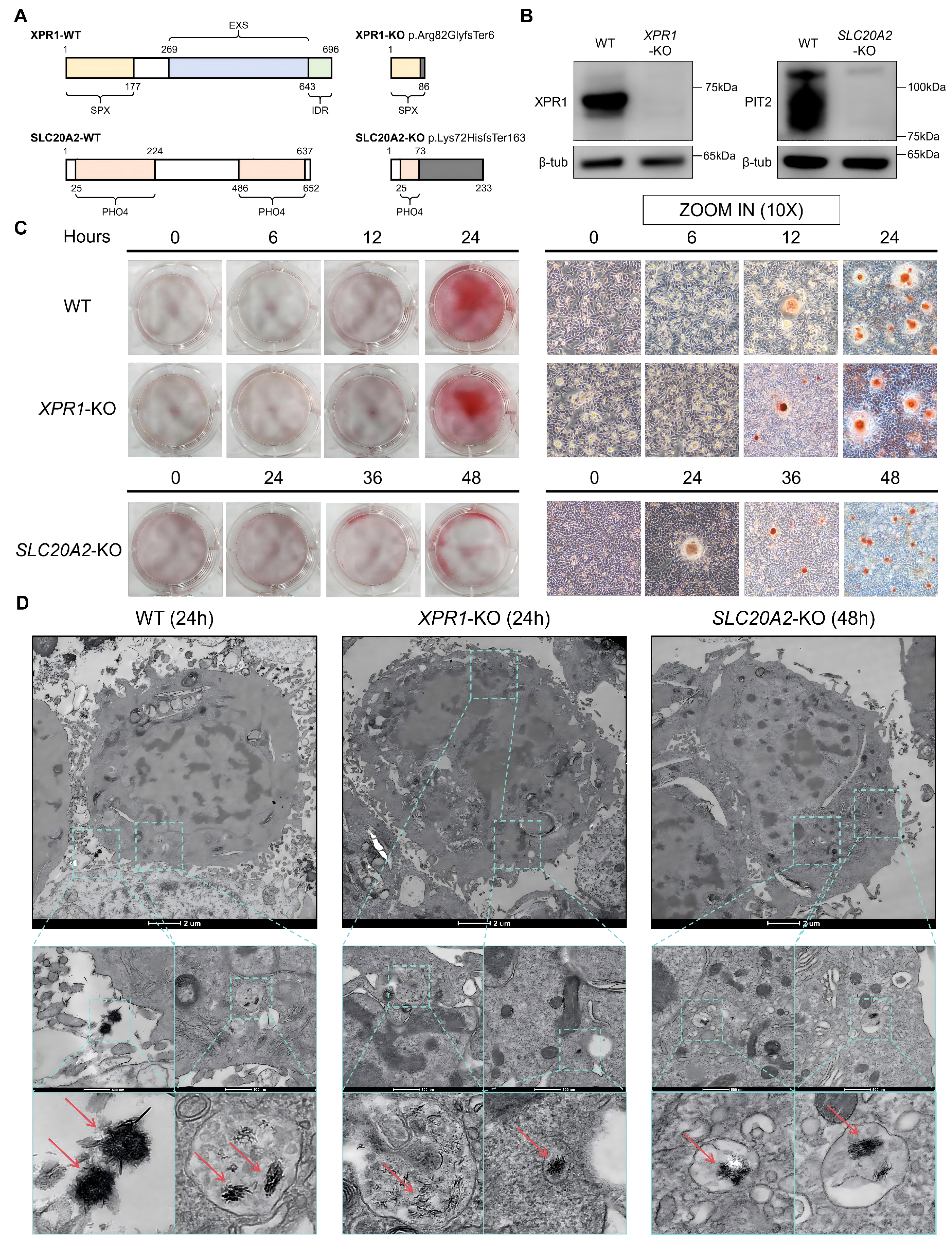
Figure 1
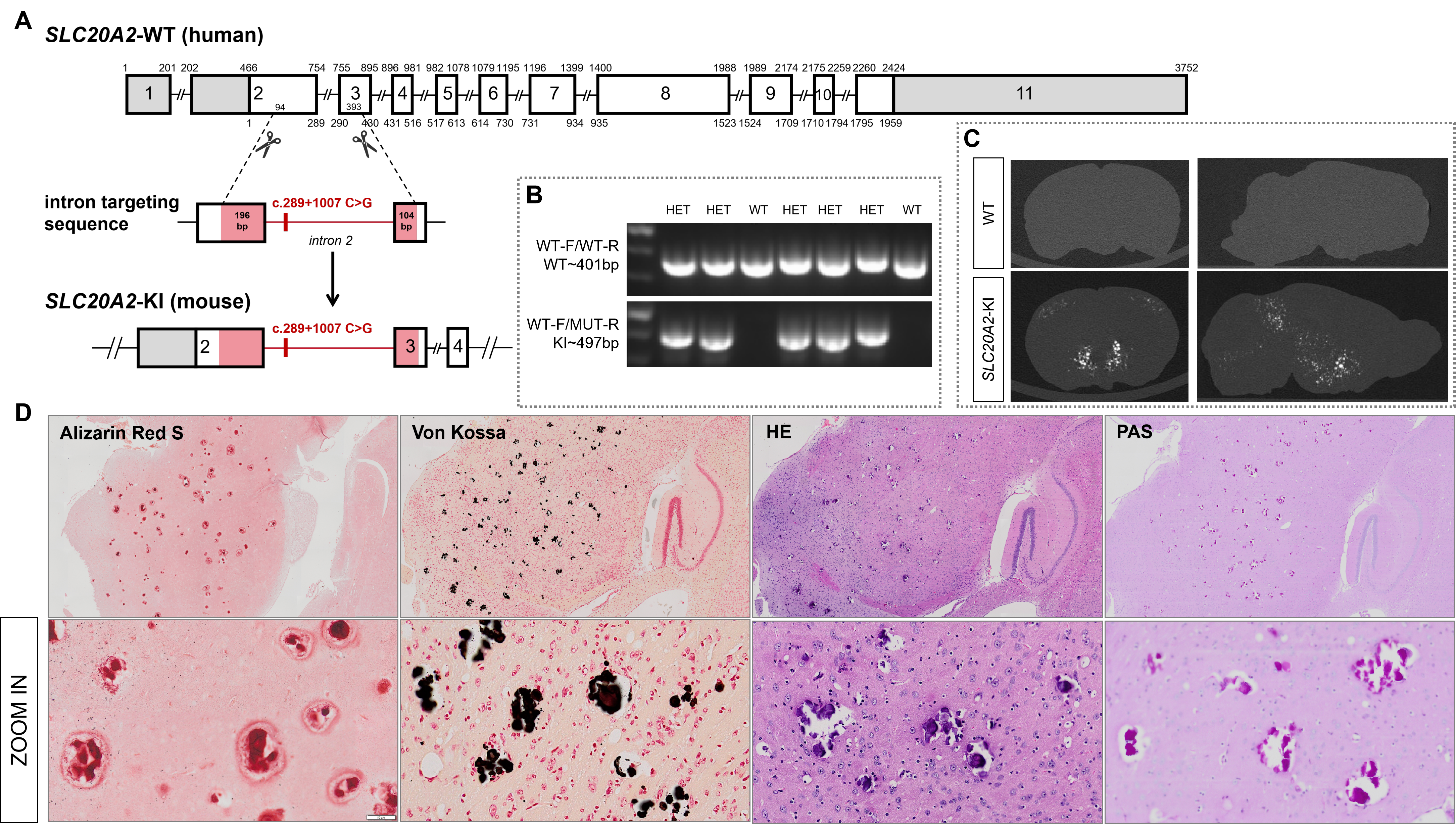
Figure 2
To cite this abstract in AMA style:
X. Jin, L. Wang, W. Luo. Calcification Foci Caused by Gene Defects in Primary Brain Calcification May Originate from Matrix Vesicle [abstract]. Mov Disord. 2026; 41 (suppl 1). https://www.mdsabstracts.org/abstract/calcification-foci-caused-by-gene-defects-in-primary-brain-calcification-may-originate-from-matrix-vesicle/. Accessed October 1, 2026.« Back to 2026 International Congress
MDS Abstracts - https://www.mdsabstracts.org/abstract/calcification-foci-caused-by-gene-defects-in-primary-brain-calcification-may-originate-from-matrix-vesicle/