
Category: MSA, PSP, CBS: Neuroimaging
Objective: To develop and evaluate a novel MRI-based volumetric diagnostic index using artificial intelligence–driven segmentation for differentiating progressive supranuclear palsy (PSP) from Parkinson’s disease (PD).
Background: Differentiating PSP from PD in early disease stages is clinically challenging, particularly for the PSP-parkinsonism (PSP-P) subtype, which often mimics PD. Conventional MRI markers such as MRPI 2.0 rely on planimetric measurements and may not fully capture three-dimensional structural brain changes. Volumetric MRI analysis combined with automated segmentation may provide more comprehensive structural information and improve diagnostic accuracy.
Method: We retrospectively analyzed brain MRI data from 127 PD patients and 128 PSP patients (including 89 PSP-Richardson syndrome and 39 PSP-parkinsonism) with disease duration <4 years. Six brain structures relevant to parkinsonian syndromes were automatically segmented using a 3D UNETR deep learning model: midbrain, pons, superior cerebellar peduncle (SCP), middle cerebellar peduncle (MCP), third ventricle, and lateral ventricles. Based on volumetric measurements, five novel diagnostic indices and volumetric adaptations of MRPI and MRPI 2.0 were developed. Diagnostic performance was evaluated using AUC, sensitivity, specificity, F1-score, and accuracy with 5-fold cross-validation and holdout validation.
Results: The automated segmentation model demonstrated high reliability with mean Dice similarity coefficients of 0.90 in PD and 0.89 in PSP. Among the evaluated indices, Index #5 (incorporating midbrain, SCP, and third ventricle volumes) showed the best diagnostic performance for distinguishing PD from PSP with AUC 0.953, sensitivity 0.887, specificity 0.924, and accuracy 0.906, outperforming conventional MRPI 2.0 in AUC, sensitivity, F1-score, and overall accuracy. The proposed index also demonstrated strong performance in subgroup analyses differentiating PD from PSP-P and PSP-RS.
Conclusion: A novel AI-driven volumetric MRI index based on automated segmentation of key brain structures improves the differentiation of PSP from PD compared with conventional planimetric approaches. This method provides a fully automated and clinically applicable imaging biomarker and may facilitate earlier and more accurate diagnosis of parkinsonian syndromes.
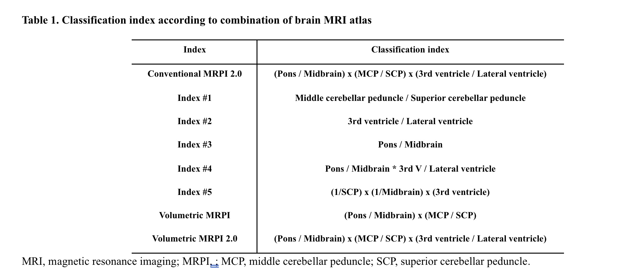
Table 1. Classification index MRI atlas
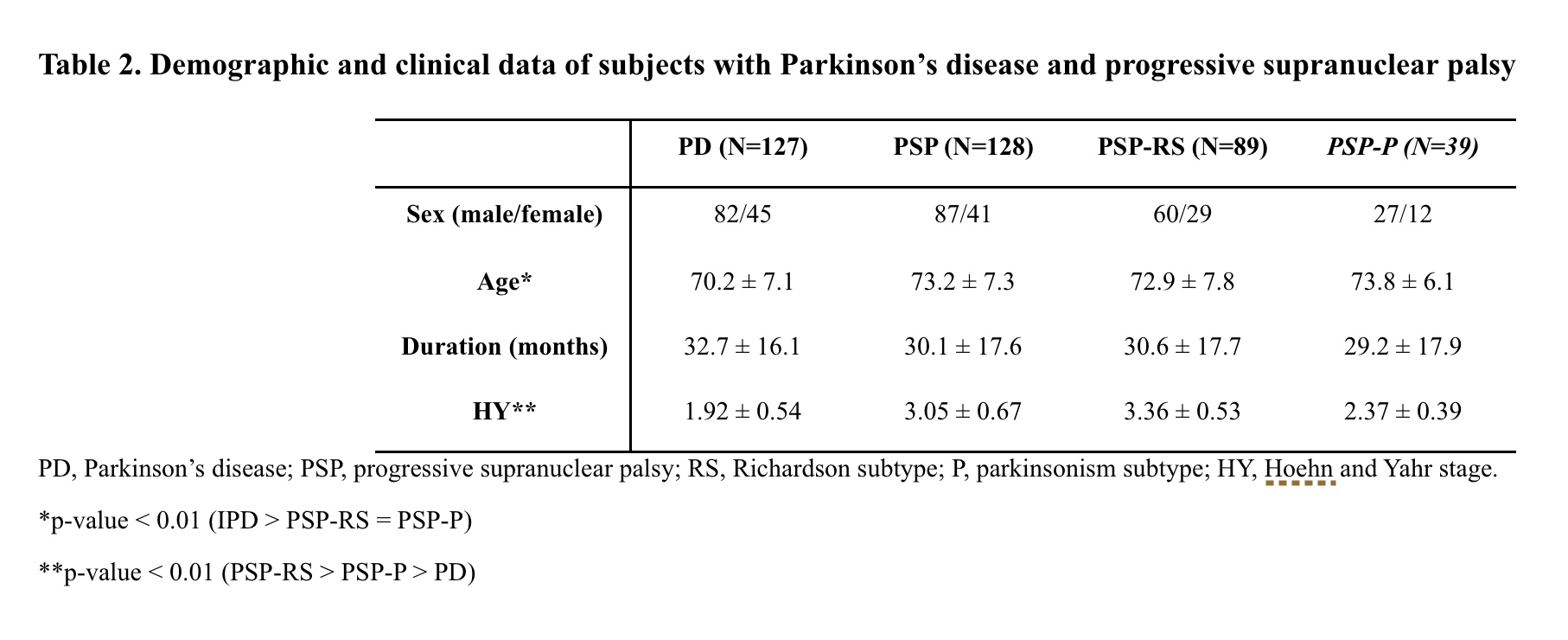
Table 2. Clinical data of subjects
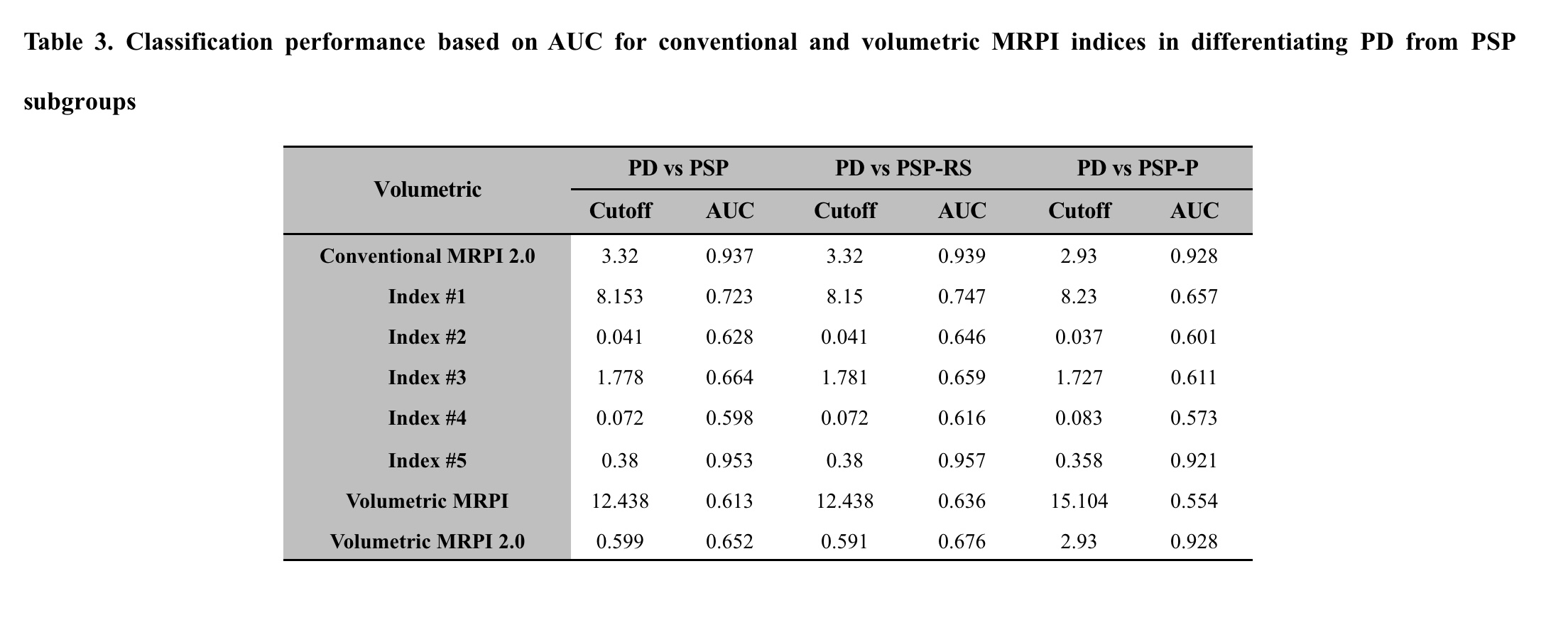
Table 3. Classification performance
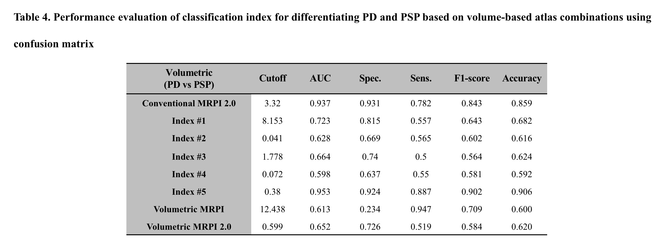
Table 4. Performance evaluation
References: This research was supported by the Korea Health Technology R&D Project through the Korean Health Industry Development Institute (KHIDI), funded by the Ministry of Health & Welfare, Republic of Korea (grant number : RS-2023-00266288).
References
1. Hughes AJ, Daniel SE, Kilford L, Lees AJ. Accuracy of clinical diagnosis of idiopathic Parkinson’s disease: a clinico-pathological study of 100 cases. J Neurol Neurosurg Psychiatry 1992;55(3):181–184.
2. Srulijes K, Mallien G, Bauer S, et al. In vivo comparison of Richardson’s syndrome and progressive supranuclear palsy-parkinsonism. J Neural Transm (Vienna) 2011;118(8):1191–1197.
3. Ahn JH, Kim M, Kim JS, et al. Midbrain atrophy in patients with presymptomatic progressive supranuclear palsy-Richardson’s syndrome. Parkinsonism Relat Disord 2019;66:80–86.
4. Madetko N, Alster P, Kutyłowski M, et al. Is MRPI 2.0 more useful than MRPI and M/P ratio in differential diagnosis of PSP-P with other atypical Parkinsonisms? Journal of Clinical Medicine 2022;11(10):2701.
5. Nigro S, Filardi M, Tafuri B, et al. Deep Learning-based Approach for Brainstem and Ventricular MR Planimetry: Application in Patients with Progressive Supranuclear Palsy. Radiol Artif Intell 2024;6(3):e230151.
6. Sun F, Lyu J, Jian S, Qin Y, Tang X. Accurate measurement of magnetic resonance parkinsonism index by a fully automatic and deep learning quantification pipeline. Eur Radiol 2023;33(12):8844–8853.
7. Sjostrom H, Granberg T, Hashim F, Westman E, Svenningsson P. Automated brainstem volumetry can aid in the diagnostics of parkinsonian disorders. Parkinsonism Relat Disord 2020;79:18–25.
8. Postuma RB, Poewe W, Litvan I, et al. Validation of the MDS clinical diagnostic criteria for Parkinson’s disease. Mov Disord 2018;33(10):1601–1608.
9. Hoglinger GU, Respondek G, Stamelou M, et al. Clinical diagnosis of progressive supranuclear palsy: The movement disorder society criteria. Mov Disord 2017;32(6):853–864.
10. Goetz CG, Tilley BC, Shaftman SR, et al. Movement Disorder Society-sponsored revision of the Unified Parkinson’s Disease Rating Scale (MDS-UPDRS): scale presentation and clinimetric testing results. Mov Disord 2008;23(15):2129–2170.
11. Chakraborty S, Aich S, Kim HC. 3D Textural, Morphological and Statistical Analysis of Voxel of Interests in 3T MRI Scans for the Detection of Parkinson’s Disease Using Artificial Neural Networks. Healthcare (Basel) 2020;8(1).
12. Ya Y, Ji L, Jia Y, et al. Machine Learning Models for Diagnosis of Parkinson’s Disease Using Multiple Structural Magnetic Resonance Imaging Features. Front Aging Neurosci 2022;14:808520.
13. Gaser C, Dahnke R, Thompson PM, Kurth F, Luders E, Initiative AsDN. CAT: a computational anatomy toolbox for the analysis of structural MRI data. GigaScience 2024;13:giae049.
14. Ashburner J, Barnes G, Chen C-C, et al. SPM12 manual. Wellcome Trust Centre for Neuroimaging, London, UK 2014;2464(4).
15. Mori S. MRI atlas of human white matter: Elsevier, 2005.
16. Hammers A, Allom R, Koepp MJ, et al. Three‐dimensional maximum probability atlas of the human brain, with particular reference to the temporal lobe. Human brain mapping 2003;19(4):224–247.
17. Shattuck DW, Mirza M, Adisetiyo V, et al. Construction of a 3D probabilistic atlas of human cortical structures. Neuroimage 2008;39(3):1064–1080.
18. Hatamizadeh A, Tang Y, Nath V, et al. Unetr: Transformers for 3d medical image segmentation. In: Proceedings of the IEEE/CVF winter conference on applications of computer vision; 2022; 2022. p. 574–584.
19. Cui SS, Ling HW, Du JJ, et al. Midbrain/pons area ratio and clinical features predict the prognosis of progressive Supranuclear palsy. BMC Neurol 2020;20(1):114.
20. Choi SM, Kim BC, Nam TS, et al. Midbrain atrophy in vascular Parkinsonism. Eur Neurol 2011;65(5):296–301.
21. Massey LA, Jager HR, Paviour DC, et al. The midbrain to pons ratio: a simple and specific MRI sign of progressive supranuclear palsy. Neurology 2013;80(20):1856–1861.
22. Quattrone A, Morelli M, Nigro S, et al. A new MR imaging index for differentiation of progressive supranuclear palsy-parkinsonism from Parkinson’s disease. Parkinsonism Relat Disord 2018;54:3–8.
23. Ota M, Sato N, Takahashi Y, et al. Correlation between the regional brain volume and glymphatic system activity in progressive supranuclear palsy. Dement Geriatr Cogn Disord 2023.
24. Hoglinger GU, Schope J, Stamelou M, et al. Longitudinal magnetic resonance imaging in progressive supranuclear palsy: A new combined score for clinical trials. Mov Disord 2017;32(6):842–852.
25. Josephs KA, Xia R, Mandrekar J, et al. Modeling trajectories of regional volume loss in progressive supranuclear palsy. Mov Disord 2013;28(8):1117–1124.
26. Koizumi R, Akagi A, Riku Y, et al. Clinicopathological features of progressive supranuclear palsy with asymmetrical atrophy of the superior cerebellar peduncle. Neuropathology 2023;43(3):233–243.
27. Tsuboi Y, Slowinski J, Josephs KA, Honer WG, Wszolek ZK, Dickson DW. Atrophy of superior cerebellar peduncle in progressive supranuclear palsy. Neurology 2003;60(11):1766–1769.
28. Nicoletti G, Caligiuri ME, Cherubini A, et al. A Fully Automated, Atlas-Based Approach for Superior Cerebellar Peduncle Evaluation in Progressive Supranuclear Palsy Phenotypes. AJNR Am J Neuroradiol 2017;38(3):523–530.
29. Whitwell JL, Master AV, Avula R, et al. Clinical correlates of white matter tract degeneration in progressive supranuclear palsy. Arch Neurol 2011;68(6):753–760.
30. Paviour DC, Price SL, Stevens JM, Lees AJ, Fox NC. Quantitative MRI measurement of superior cerebellar peduncle in progressive supranuclear palsy. Neurology 2005;64(4):675–679.
To cite this abstract in AMA style:
MS. Cho, JY. Youn, JW. Cho, HJ. Yoo, SJ. Oh, JH. Ahn, JW. Yu, MJ. Chung. Volumetric Imaging Diagnostic Tool to Differentiate Progressive Supranuclear Palsy from Parkinson’s Disease using Artificial Intelligence [abstract]. Mov Disord. 2026; 41 (suppl 1). https://www.mdsabstracts.org/abstract/volumetric-imaging-diagnostic-tool-to-differentiate-progressive-supranuclear-palsy-from-parkinsons-disease-using-artificial-intelligence/. Accessed October 1, 2026.« Back to 2026 International Congress
MDS Abstracts - https://www.mdsabstracts.org/abstract/volumetric-imaging-diagnostic-tool-to-differentiate-progressive-supranuclear-palsy-from-parkinsons-disease-using-artificial-intelligence/