
Category: Parkinson's disease: Neuroimaging
Objective: To identify rs-fMRI-derived functional brain network patterns associated with parkinsonian tremor severity and dopaminergic medication responsiveness in Parkinson’s disease (PD).
Background: While metabolic imaging studies have identified PD tremor-related disease networks, the functional brain networks associated with tremor severity and dopa-responsiveness have not been fully characterized using rs-fMRI.
Method: rs-fMRI data were acquired from 148 PD patients (mean age 60.3 ± 9.0 years, 86 males) and 37 healthy controls (HC, 58.2 ± 5.8 years, 13 females). Patients were randomly divided into a training set (n = 74) and an independent testing set (n = 74). Group-ICA was used and functional PD-related pattern (fPDRP) distinguishing PD patients from HC was identified using logistic regression in the training set and validated in the testing set. Linear regression analyses were used to derive a functional PD tremor-related pattern (fPDTP) associated with tremor severity. Finally, an exploratory regression analysis was conducted to identify a functional PD tremor dopa-responsiveness-related pattern (fPDTRP) associated with tremor improvement following dopaminergic treatment.
Results: The fPDRP reliably discriminated PD patients from HC and was characterized by prominent contributions from thalamo-striatal circuitry, the frontoparietal network, cerebellum, temporo-parietal association cortex, and limbic regions [Figure1,2]. The fPDTP was characterized by a spatial covariance profile involving the frontoparietal network, thalamo-striatal regions, temporo-occipital association cortex, cerebellum, salience network, and orbitofrontal cortex. fPDTP expression showed significant correlations with tremor severity in the testing set (Spearman’s rho = 0.27, p = 0.01) and was significantly higher in tremor-dominant (TD) patients than in PIGD patients (p = 0.008) [Figure3]. An exploratory analysis identified an fPDTRP involving the motor cortex, frontoparietal network, and orbitofrontal cortex. Expression of this pattern correlated with the degree of tremor improvement following dopaminergic medication (Spearman’s rho = 0.29, p = 0.001) and differed significantly between dopa-responsive and dopa-resistant TD patients (p = 0.003) [Figure4].
Conclusion: These findings indicate that parkinsonian tremor and its dopa-responsiveness are mediated by distinct functional brain networks that can be identified using rs-fMRI.
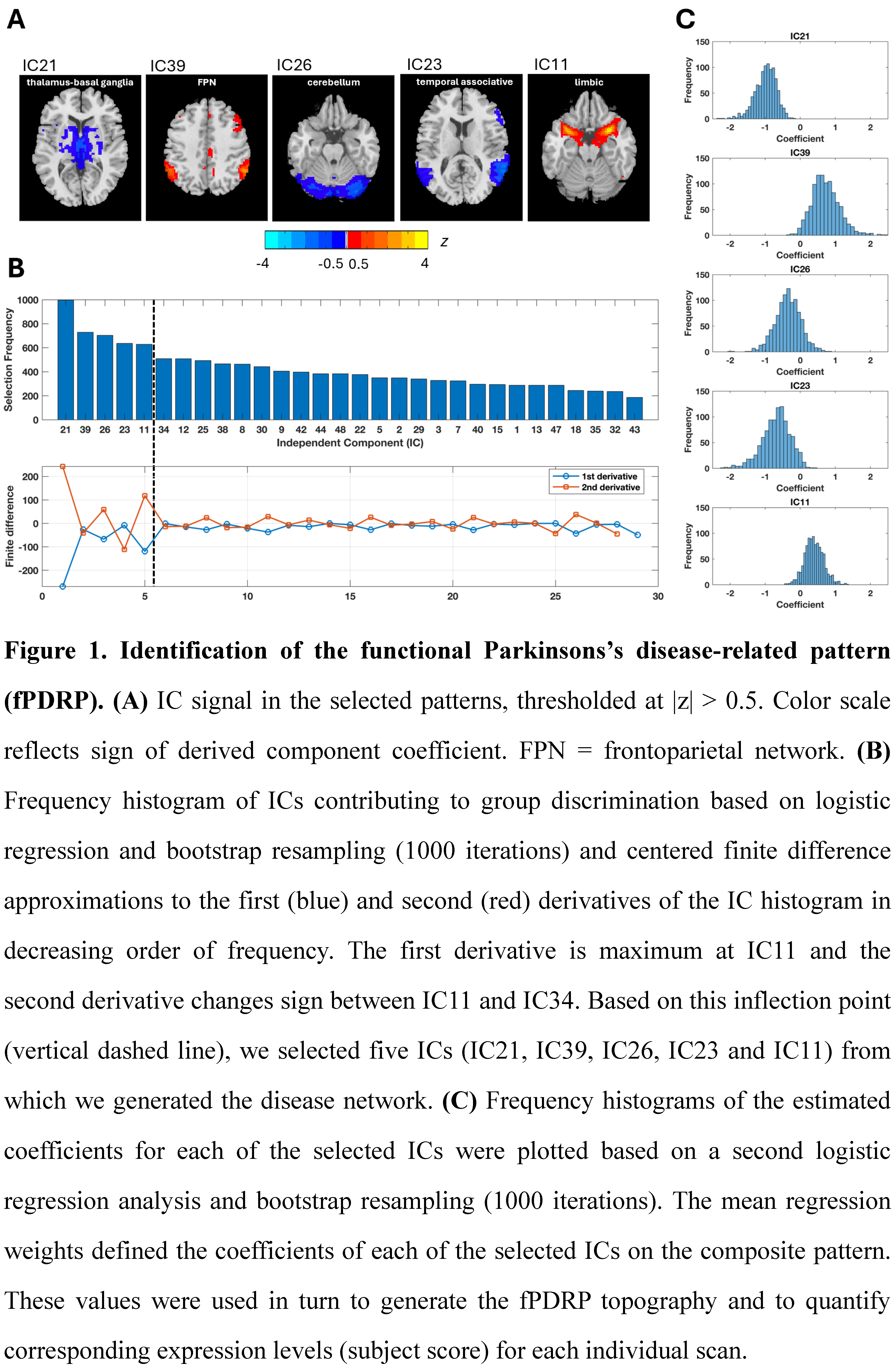
Figure 1
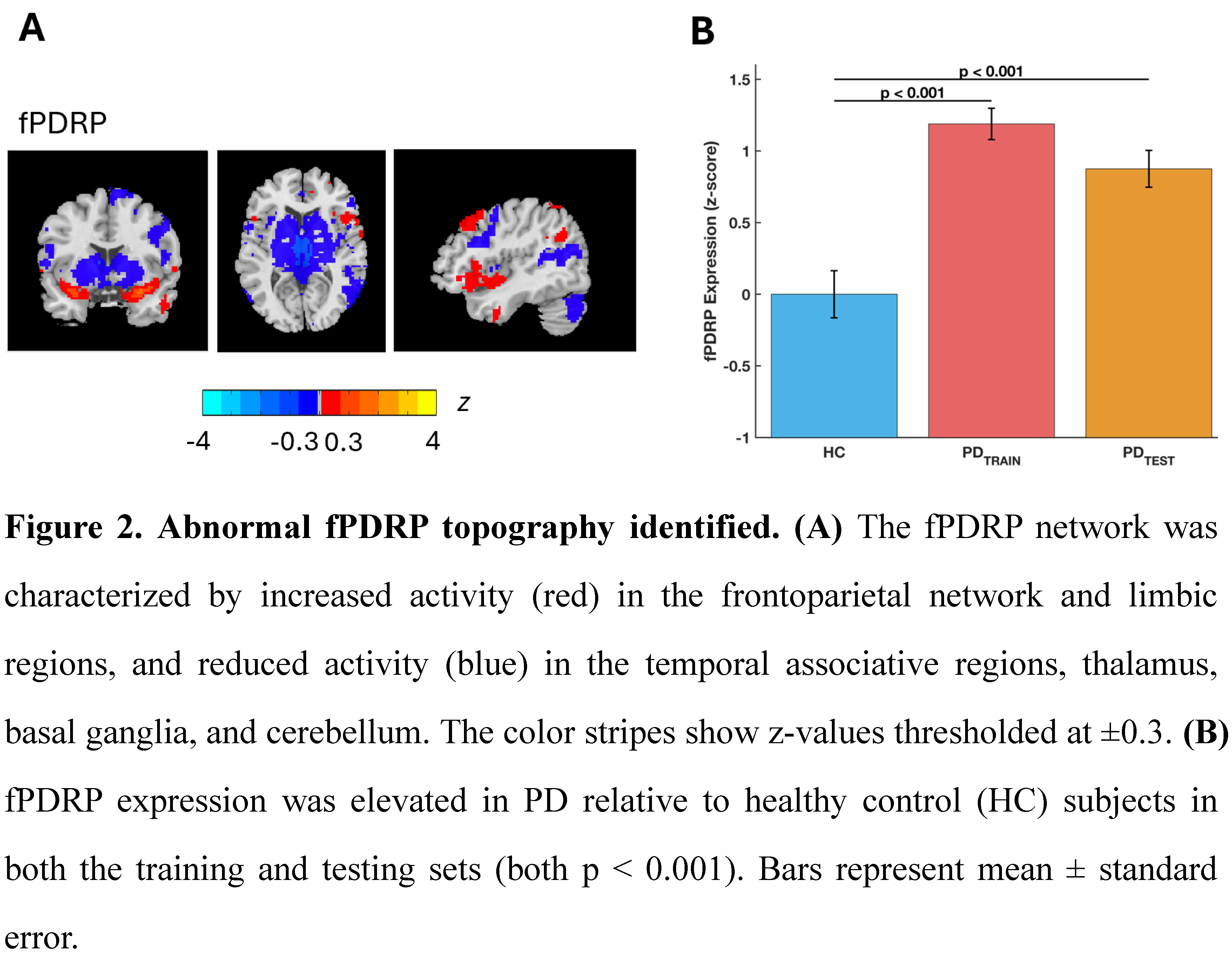
Figure 2
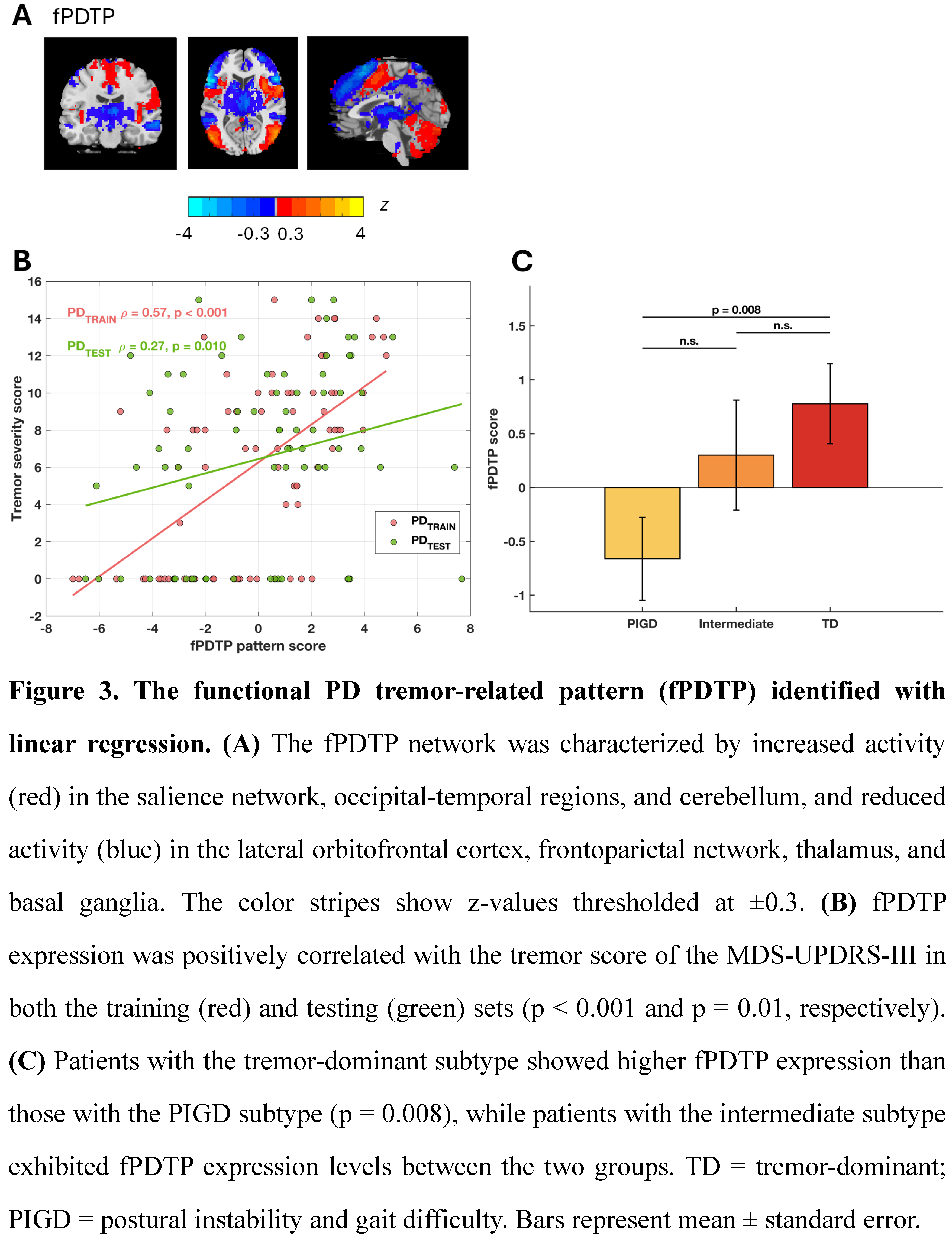
Figure 3
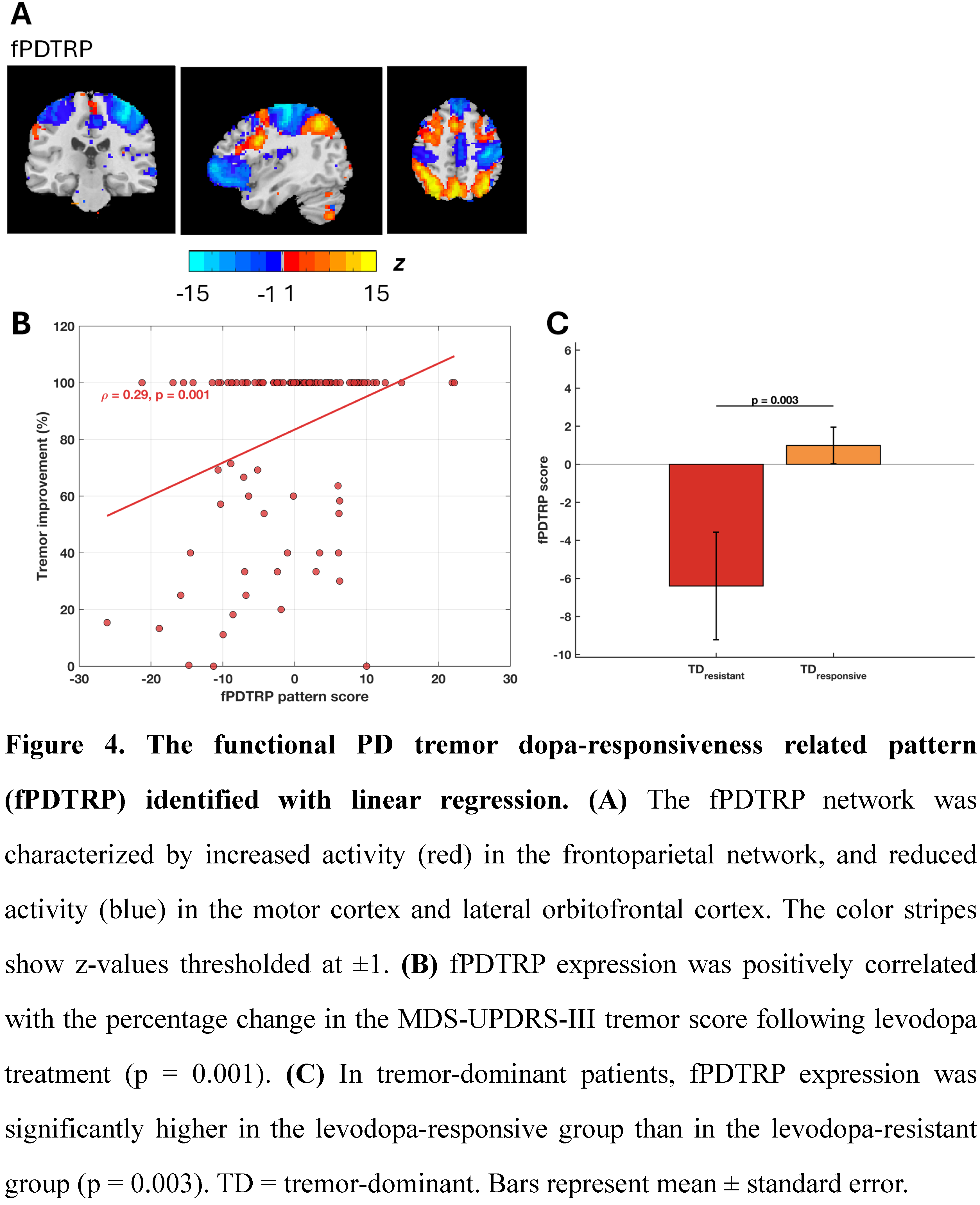
Figure 4
References: [1] Ma Y, Tang C, Spetsieris PG, Dhawan V, Eidelberg D. Abnormal metabolic network activity in Parkinson’s disease: test-retest reproducibility. J Cereb Blood Flow Metab. 2007 Mar;27(3):597-605.
[2] Mure H, Hirano S, Tang CC, Isaias IU, Antonini A, Ma Y, Dhawan V, Eidelberg D. Parkinson’s disease tremor-related metabolic network: characterization, progression, and treatment effects. Neuroimage. 2011 Jan 15;54(2):1244-53.
[3] Vo A, Sako W, Fujita K, Peng S, Mattis PJ, Skidmore FM, Ma Y, Uluğ AM, Eidelberg D. Parkinson’s disease-related network topographies characterized with resting state functional MRI. Hum Brain Mapp. 2017 Feb;38(2):617-630.
[4] Ellis EG, Meyer GM, Kaasinen V, Corp DT, Pavese N, Reich MM, Joutsa J. Multimodal neuroimaging to characterize symptom-specific networks in movement disorders. NPJ Parkinsons Dis. 2024 Aug 14;10(1):154.
To cite this abstract in AMA style:
ZY. Lin, J. Li, ZT. Zeng, DY. Li. Identification of Parkinson’s Disease Tremor-Related Functional Network Topographies Using 3.0 T rs-fMRI [abstract]. Mov Disord. 2026; 41 (suppl 1). https://www.mdsabstracts.org/abstract/identification-of-parkinsons-disease-tremor-related-functional-network-topographies-using-3-0-t-rs-fmri/. Accessed October 1, 2026.« Back to 2026 International Congress
MDS Abstracts - https://www.mdsabstracts.org/abstract/identification-of-parkinsons-disease-tremor-related-functional-network-topographies-using-3-0-t-rs-fmri/