
Category: Parkinsonism (Other)
Objective: The aim of this study was to describe a series of patients with brain calcifications within the Fahr spectrum and to analyze their clinical–radiological correlations
Background: The Fahr spectrum includes Fahr’s disease (FD), a primary genetic disorder, and Fahr’s syndrome (FS), caused by secondary factors. Distinguishing them is clinically important due to differences in prognosis, treatment, and genetic counseling. Both are rare, with a prevalence of less than one case per million
Method: A retrospective analysis was conducted of patients evaluated between 2018 and 2025 at a tertiary hospital in Buenos Aires, Argentina. Patients with extensive supra- and infratentorial brain calcifications identified on computed tomography (CT) or magnetic resonance imaging (MRI), with or without neurological manifestations, evaluated in movement disorders and endocrinology clinics were included. Physiological calcifications were excluded. Demographic data, clinical manifestations, neuroimaging findings, and treatments were recorded. Calcifications were classified according to their neuroimaging distribution as cortical gray matter, deep gray nuclei, subcortical white matter, and cerebellum/brainstem
Results: Six patients were identified (F:5/M:1) with a mean age of 43 years. Only one patient was asymptomatic; the mean age at symptom onset was 38 years. All patients exhibited parkinsonism. In patients with FD manifestations included epilepsy (n=3), psychiatric symptoms (n=2), chorea (n=1), cognitive impairment (n=1), ataxia (n=1), and dystonia (n=1).
Neuroimaging showed calcifications in the deep gray nuclei in all patients. Two patients showed additional involvement of the subcortical white matter, while four presented more extensive involvement that included subcortical white matter, cortical gray matter, cerebellum and brainstem
Conclusion: The involvement of the basal ganglia was associated with the presence of parkinsonism in all patients. There is no correlation between the topography and extent of calcifications and the presence or severity of symptoms. Patients with FD had a higher frequency and greater severity of symptoms than those with FS. This series highlights the importance of an adequate etiological evaluation, considering the heterogeneity of clinical presentation, the need for family risk counseling, and the prognosis of both entities
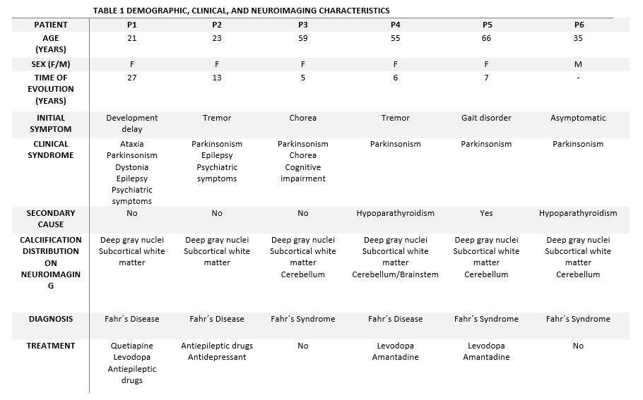
Clinical, Demographic, and Neuroimaging Data
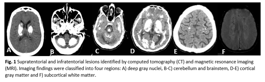
Supratentorial and Infratentorial Lesions (CT/MRI)
References: Saleem S, Aslam HM, Anwar M, Anwar S, Saleem M, Saleem A, Rehmani MAK. Fahr’s syndrome: literature review of current evidence. Orphanet J Rare Dis. 2013;8:156. doi:10.1186/1750-1172-8-156.
Saleh S, Kamourieh S, Vandrovcova J, Khan A. The spectrum of Fahr’s disease. NPJ Parkinsons Dis. 2020;6:26. doi:10.1038/s41531-020-00116-8.
Manyam BV. What is and what is not ‘Fahr’s disease’. Parkinsonism Relat Disord.
2005;11(2):73–80. doi:10.1016/j.parkreldis.2004.06.007
To cite this abstract in AMA style:
A. Ortiz Cardenas, F. Ramos, S. Rodriguez Quiroga, R. Piccioni, A. Tettamanti, M. Drnovsek, M. Nogueira, N. Garretto, T. Arakaki. Clinical–Radiological Correlation in Patients with brain calcifications: a case series [abstract]. Mov Disord. 2026; 41 (suppl 1). https://www.mdsabstracts.org/abstract/clinical-radiological-correlation-in-patients-with-brain-calcifications-a-case-series/. Accessed October 1, 2026.« Back to 2026 International Congress
MDS Abstracts - https://www.mdsabstracts.org/abstract/clinical-radiological-correlation-in-patients-with-brain-calcifications-a-case-series/