
Category: MSA, PSP, CBS: Neuroimaging
Objective: To compare absolute versus ratio brainstem planimetric measures on routine 1.5T MRI and to identify the best-performing metrics for differentiating PSP and MSA in a Vietnamese cohort.
Background: Differentiating PSP and MSA from Parkinson’s disease (PD) is difficult early in disease. Brainstem planimetry may provide practical diagnostic support, but evidence from routine 1.5T clinical scans in Asian populations is limited.
Method: In this prospective study (June 2022–January 2024), 35 participants (PSP n=5, MSA n=10, PD n=10, controls n=10) underwent 1-mm isotropic T1-weighted 1.5T MRI. A blinded neurologist measured midbrain area (MA), pons area (PA), superior cerebellar peduncle (SCP) width, middle cerebellar peduncle (MCP) width, and derived indices (MPAR, MPR, MRPI) using ImageJ. Group differences were tested with Kruskal–Wallis and Dunn’s tests, with epsilon-squared (ε²) as effect size. Diagnostic performance (AUC, sensitivity, specificity) was estimated with bootstrap 95% confidence intervals.
Results: Ratio indices showed larger group effects than absolute measures, with strongest separation for MPR (ε²=0.569; p<0.001), followed by MRPI (ε²=0.403; p=0.001). Among single measures, MCP width showed the largest effect (ε²=0.445; p=0.001), while SCP width did not differ between groups (p=0.765) (Figure 1). For predefined classifications, MPR discriminated PSP vs non-PSP (AUC=0.987 with 95% CI [0.956-1.000]; cut-off 0.48; sensitivity 100%; specificity 96.67% with 95%CI [90-100]), and MRPI discriminated MSA vs non-MSA (AUC=0.924 with 95% CI [83.71-100]; cut-off 10.16; sensitivity 100%; specificity 72% with 95% CI [56-88]) (Figure 2).
Conclusion: On routine 1.5T MRI, manual brainstem planimetry—particularly MPR for PSP and MRPI/MCP-based measures for MSA—provides feasible, low-cost diagnostic support in settings without advanced imaging or automated segmentation.
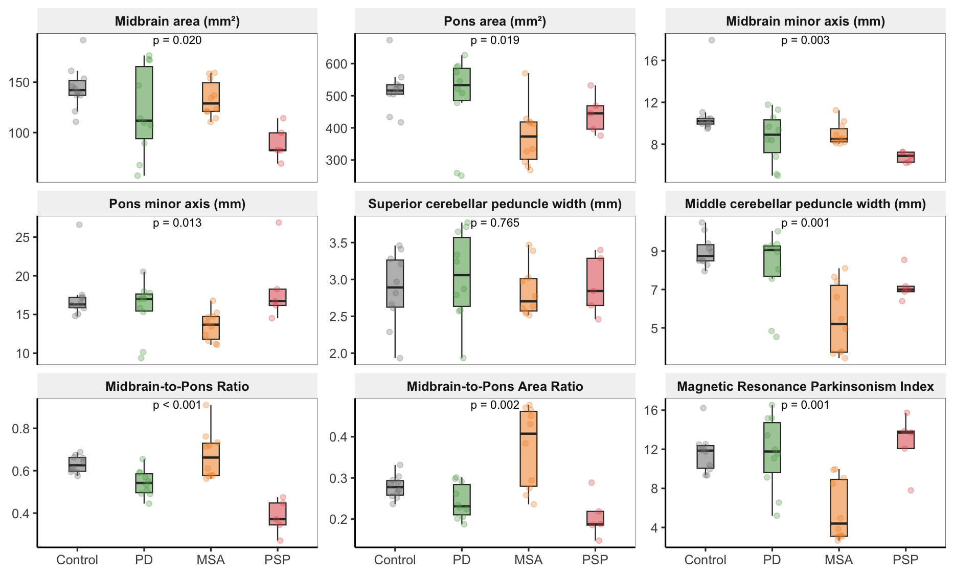
Figure 1. Brainstem planimetrics across diagnoses
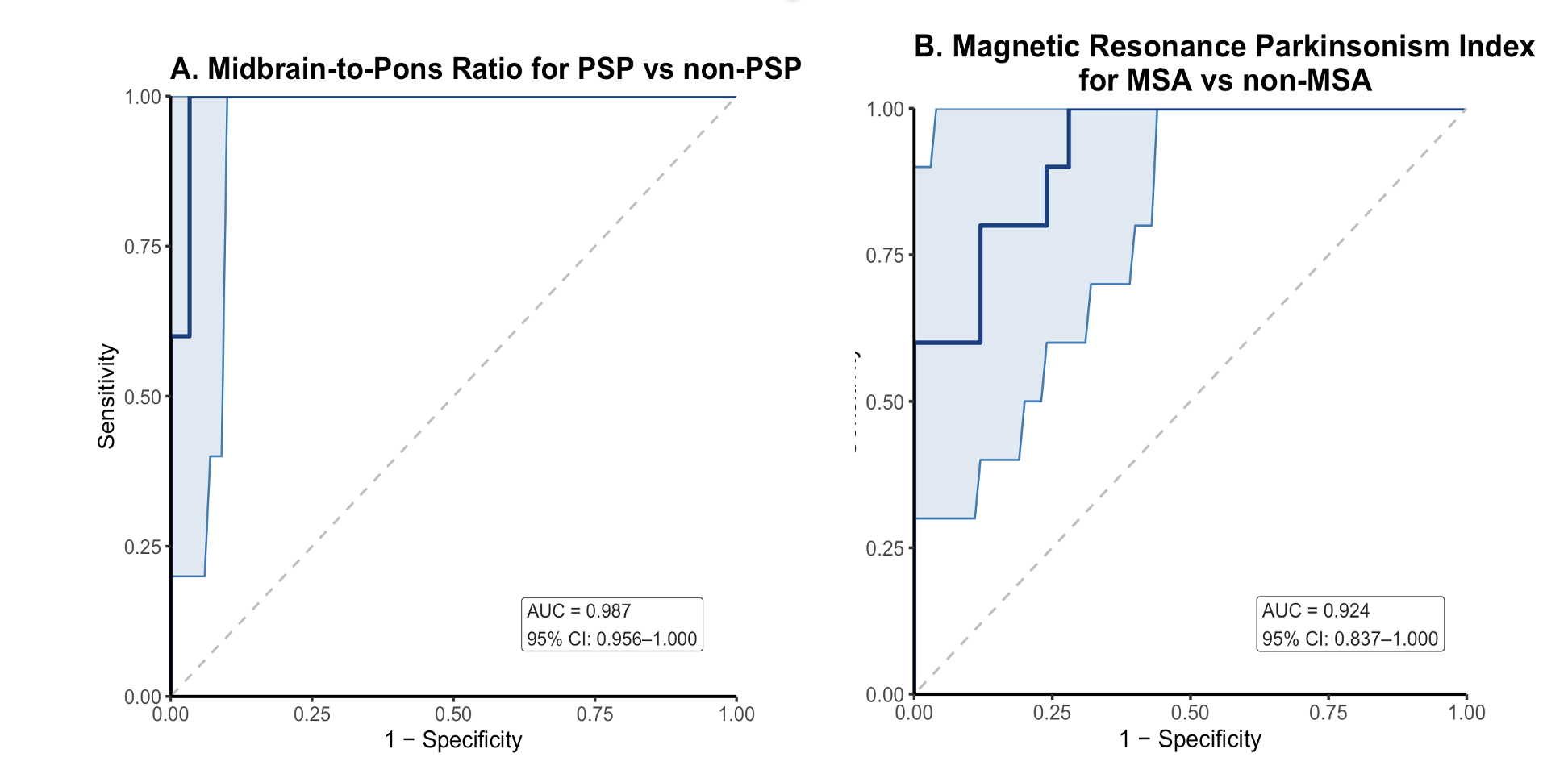
Figure 2. ROC curves (A) PSP (B) MSA vs. others
To cite this abstract in AMA style:
D. Nguyen, T. Tran. Diagnostic Utility of Routine 1.5T Brainstem Planimetry in Parkinsonism: Evidence from a Vietnamese Cohort [abstract]. Mov Disord. 2026; 41 (suppl 1). https://www.mdsabstracts.org/abstract/diagnostic-utility-of-routine-1-5t-brainstem-planimetry-in-parkinsonism-evidence-from-a-vietnamese-cohort/. Accessed October 1, 2026.« Back to 2026 International Congress
MDS Abstracts - https://www.mdsabstracts.org/abstract/diagnostic-utility-of-routine-1-5t-brainstem-planimetry-in-parkinsonism-evidence-from-a-vietnamese-cohort/