
Category: Parkinson's disease: Neuroimaging
Objective: The aim of this study is to compare the right and left vagus nerve (VN) cross-sectional area (CSA) and autonomic symptoms in patients with Parkinson’s disease (PD) carrying GBA1 gene variants (GBA-PD), GBA-negative PD patients, and healthy controls (HC).
Background: Autonomic dysfunction is highly prevalent in GBA-PD, a condition associated with variants in the gene encoding the glucocerebrosidase enzyme [1][2][3][4][5]. In α-synucleinopathies, the VN is a well-recognized site of neurodegeneration [6][7]. Although recent evidence using ultrasonography (US) has highlighted VN atrophy in PD patients [8][9][10], results remain conflicting [11][12]. Notably, most available data are based on clinical diagnosis or motor symptoms, while the specific impact of the genetic background on VN morphology has not been fully addressed.
Method: This pilot study included 16 GBA-PD patients, 15 GBA-negative PD patients, and 25 HC. VN CSA was assessed bilaterally at the level of the thyroid isthmus using ultrasonography. Clinical data [table1] and dysautonomia questionnaires were collected to explore correlations with VN morphological measurements.
Results: Preliminary data based exclusively on VN CSA demonstrated that total mean VN CSA was significantly lower in GBA-PD (1.29 ± 0.33 mm²) compared to both GBA-negative PD (2.00 ± 0.34 mm², p < 0.001) and HC (2.13 ± 0.38 mm², p < 0.001). In GBA-PD VN CSA measured 1.32 ± 0.36 mm² (right) and 1.26 ± 0.32 mm² (left). In GBA-negative PD VN CSA measured 2.09 ± 0.38 mm² (right) and 1.91 ± 0.29 mm² (left); HC showed values of 2.19 ± 0.38 mm² (right) and 2.03 ± 0.36 mm² (left). No significant differences were found between GBA-negative PD and HC for either the right (p=0.42) or left side (p=0.31). Physiological asymmetry (Right > Left) was confirmed in HC (p=0.015), differently from the GBA-negative PD (p=0.10) and was entirely lost in GBA-PD (p=0.53) [figure1] [table2].
Conclusion: These findings demonstrate a significant reduction of VN CSA in GBA-PD patients compared to GBA-negative PD and HC. Adopting a genotype-driven approach, we identify VN atrophy as a distinctive morphological feature of GBA-PD. The loss of physiological asymmetry suggests severe bilateral vagal nerve involvement in PD patients, supporting VN sonographic evaluation as a potential morphological biomarker, especially in the GBA-PD population.
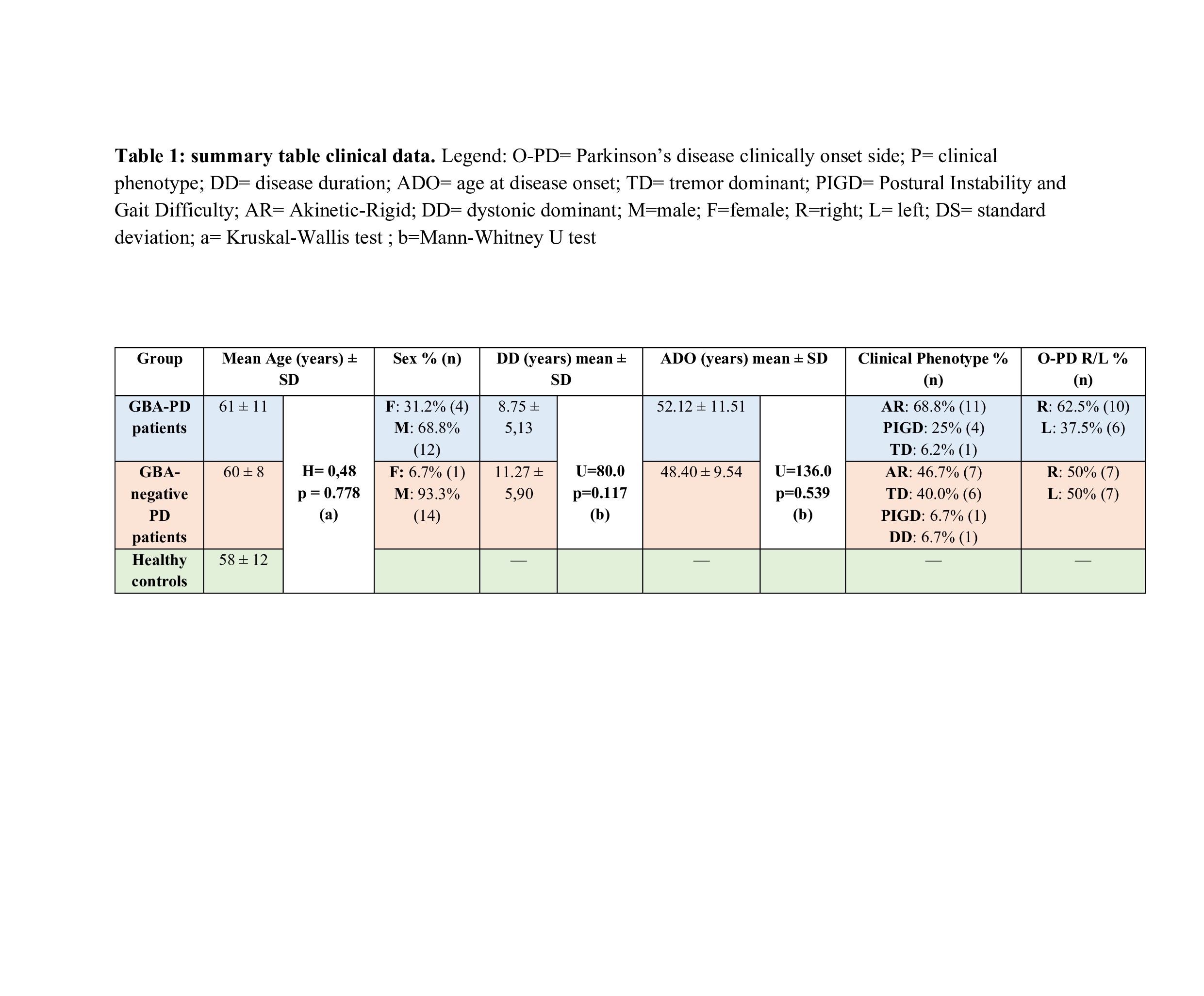
Table 1
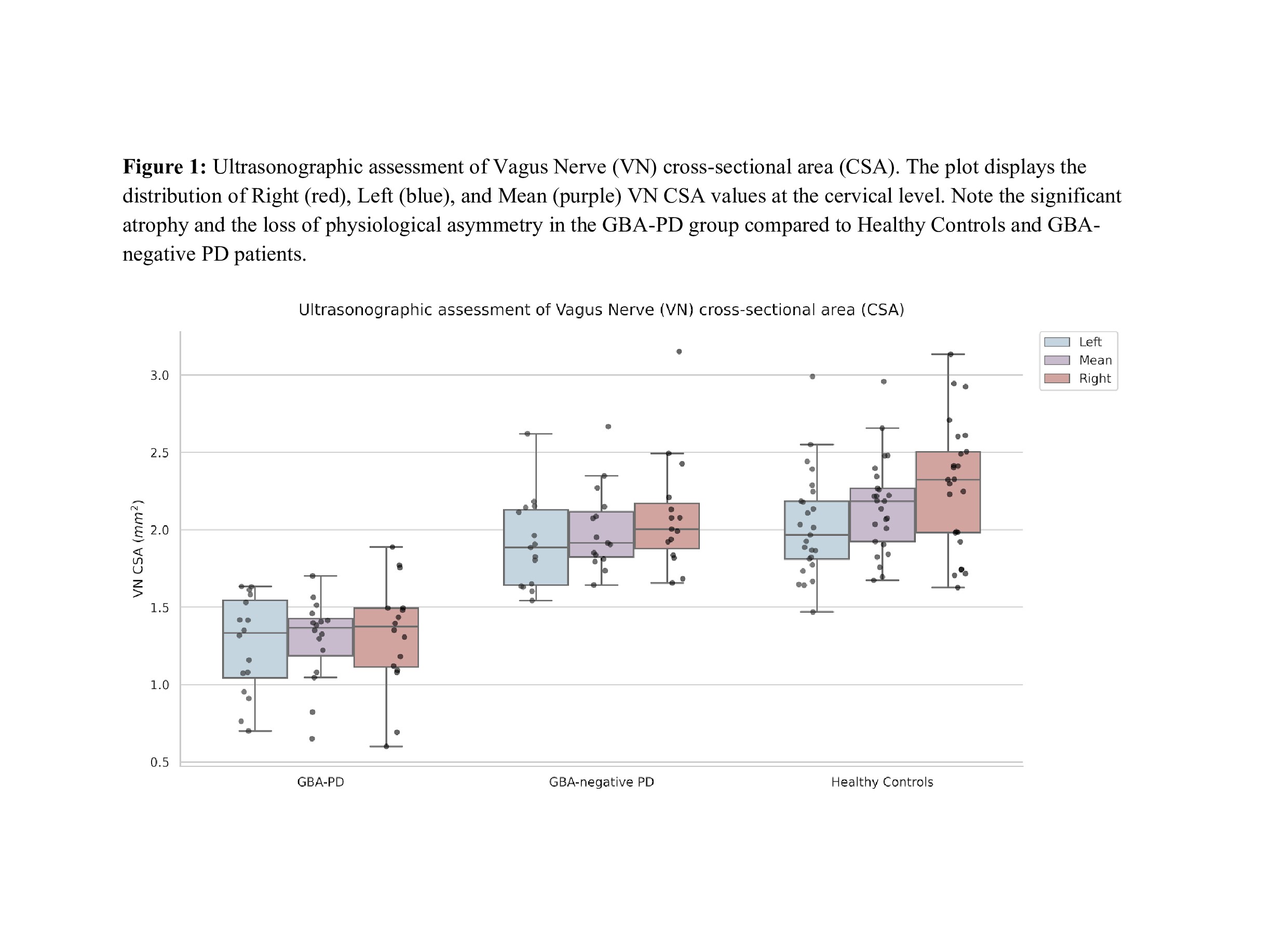
Figure 1
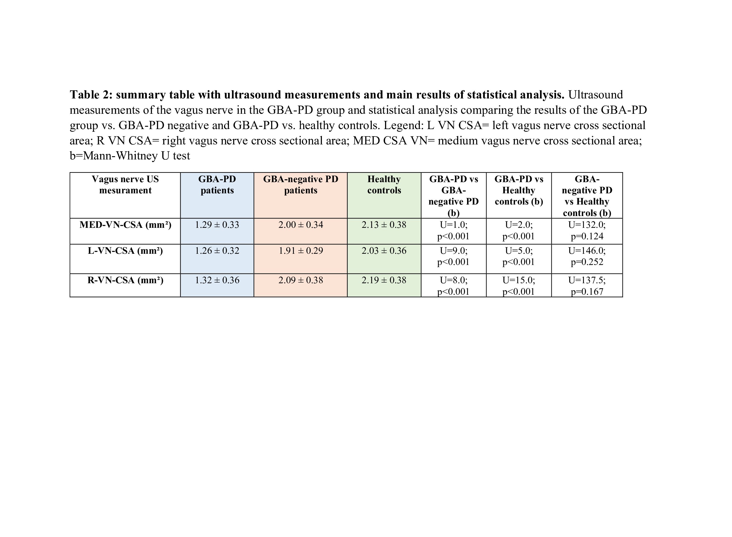
Table 2
References: 1. Zhou, Y.; Wang, Y.; Wan, J.; Zhao, Y.; Pan, H.; Zeng, Q.; Zhou, X.; He, R.; Zhou, X.; Xiang, Y.; et al. Mutational Spectrum and Clinical Features of GBA1 Variants in a Chinese Cohort with Parkinson’s Disease. NPJ Park. Dis. 2023, 9, 129. doi: 10.1038/s41531-023-00571-4.
2. Goker-Alpan, O.; Lopez, G.; Vithayathil, J.; Davis, J.; Hallett, M.; Sidransky, E. The Spectrum of Parkinsonian Manifestations Associated with Glucocerebrosidase Mutations. Arch. Neurol. 2008, 65, 1353–1357. doi: 10.1001/archneur.65.10.1353.
3. A.Carandina, G. Lazzeri, G. Dias Rodrigues, G. Franco, E. Monfrini, F. Arienti, E. Frattini, I. Trezzi, P. Paulo da Silva Soares, C. Bellocchi, L.Furlan, N.Montano, A.Di Fonzo and E.Tobaldini. Dysautonomia in Parkinson’s Disease: Impact of Glucocerebrosidase Gene Mutations on Cardiovascula r Autonomic Control. Frontiers in neuroscience. doi: 10.3389/fnins.2022.842498
4. Aliaksandr Skrahin, Mia Horowitz, Majdolen Istaiti, Volha Skrahina, Jan Lukas, Gilad Yahalom, Mikhal E. Cohen, Shoshana Revel-Vilk, Ozlem Goker-Alpan, Michal Becker-Cohen, Sharon Hassin-Baer, Per Svenningsson, Arndt Rolfs and Ari Zimran. GBA1-Associated Parkinson’s Disease Is a Distinct Entity. Int J Mol Sci. 2024 Jun 28;25(13):7102. doi: 10.3390/ijms25137102.
5. G. Devigili, G. Straccia, E. Cereda, B. Garavaglia, A. Fedeli, A. Emanuele Elia, S. H. M. J. Piacentini, S. Prioni, P. Amami, F. Invernizzi, N. G. Andreasi, L. Michele Romito, R. Eleopra, R.Cilia. Unraveling Autonomic Dysfunction in GBA-Related Parkinson’s Disease. Mov. Disord Clin Pract. 2023 Oct 13;10(11):1620-1638. doi: 10.1002/mdc3.13892. eCollection 2023 Nov.
6. David P. Breen, Glenda M. Halliday and Anthony E. Lang. Gut–Brain Axis and the Spread of α-Synuclein Pathology: Vagal Highway or Dead End? Movement Disorders : Official Journal of the Movement Disorder Society. 2019. doi: 10.1002/mds.27556.
7. R. Takahashi, H. Yamakado, N. Uemura, T. Taguchi and J. Ueda. The Gut–Brain Axis Based on α-Synuclein Propagation—Clinical, Neuropathological, and Experimental Evidence. Int J Mol Sci. 2025 Apr 23;26(9):3994.doi: 10.3390/ijms26093994.
8. Y. Cui, R. Yan, Y. Qu, S.Liu, X. Li. Ultrasonography of the vagus nerve in Parkinson’s disease: a systematic review and meta‑analysis. Neurological Sci. 2025 Jul. doi: 10.1007/s10072-025-08346-w. Epub 2025 Jul 14.
9. R. Abdelnaby, M. Elsayed, K. A. Mohamed, K. T. Dardeer, Y.T. Sonbol, A. ELgenidy, M. H. Barakat, M. M. Alwerdani, A. Maier. Vagus nerve ultrasonography in Parkinson’s disease: A systematic review and meta-analysis. Auton Neurosci. 2021 Sep:234:102835. Doi: 10.1016/j.autneu.2021.102835. Epub 2021 Jun 20.
10. R. Abdelnaby, M. H. El Din Moawad, A. S. Shabib, K. Ashraf Mohamed, M. A. Ebrahim, A. M. Aboutaleb, D. E. Gaber, A. S. Serour, M. A. Shamim, M. H. Elberry, M. A. Bedewi, Mohamed Elsayed, U. Walter. Sonographic vagus nerve atrophy in Parkinson’s disease: Myth or fact? A systematic review and meta-analysis of recent evidence answers. Parkinsonism Relat Disord. 2023 Jul:112:105451.doi: 10.1016/j.parkreldis.2023.105451. Epub 2023 May 22.
11. N. Fedtke, O. W Witte, T. Prell. Ultrasonography of the Vagus Nerve in Parkinson’s Disease. Front Neurol. 2018 Jul 2:9:525. doi: 10.3389/fneur.2018.00525. eCollection 2018.
12. J. Horsager, U. Walter, T. D. Fedorova, K. B. Andersen, C. Skjærbæk, K. Knudsen, N. Okkels, P. von Weitzel-Mudersbach, S. E. Dyrskog, B. Bergholt, P. Borghammer. Vagus Nerve Cross-Sectional Area in Patients With Parkinson’s Disease-An Ultrasound Case-Control Study. Front Neurol. 2021 Jun 22:12:681413.doi: 10.3389/fneur.2021.681413. eCollection 2021.
To cite this abstract in AMA style:
J. Di Giovanni, D. Genovese, G. Granata, G. Di Lazzaro, F. Tomasello, A. Cimmino, M. Patera, A. de Biase, M. Petracca, C. Piano, F. Bove, A. Bentivoglio, P. Calabresi. Genotype Matters: Vagus Nerve Atrophy in GBA-Associated Parkinson’s Disease [abstract]. Mov Disord. 2026; 41 (suppl 1). https://www.mdsabstracts.org/abstract/genotype-matters-vagus-nerve-atrophy-in-gba-associated-parkinsons-disease/. Accessed October 1, 2026.« Back to 2026 International Congress
MDS Abstracts - https://www.mdsabstracts.org/abstract/genotype-matters-vagus-nerve-atrophy-in-gba-associated-parkinsons-disease/