
Category: Parkinson's disease: Neuroimaging
Objective: To evaluate a deep learning-based automated segmentation pipeline for extracting midbrain regions in MRI scans related to Parkinson’s disease.
Background: Magnetic resonance imaging has become an important modality for investigating structural biomarkers in Parkinson’s disease (PD). In particular, the midbrain region is critical for studying neurodegenerative changes associated with dopaminergic neuron loss [1]. Traditional segmentation techniques rely on handcrafted rules and often struggle with low-contrast MRI images [2].
Method: MRI scans were obtained from the Parkinson’s Progression Markers Initiative dataset, including 151 Parkinson’s disease patients and 100 healthy controls. Among them, 30 scans contained expert-annotated midbrain masks for supervised learning. An Attention U-Net architecture was used to enhance feature selection by suppressing irrelevant background signals and highlighting anatomically relevant regions. Images were preprocessed through cropping, normalization, and resizing prior to GPU-accelerated training. Model evaluation employed five-fold cross-validation with metrics including Dice coefficient, precision, recall, and F1 score.
Results: The proposed model achieved a Dice coefficient of approximately 0.82, outperforming baseline CNN segmentation (Dice ≈ 0.73) and conventional U-Net models (Dice ≈ 0.78). Stable convergence across training epochs demonstrated reliable feature learning despite the limited annotated dataset. Figure 1 shows the ground truth and prediction images for Performance comparison between baseline CNN, conventional U-Net, and Attention U-Net models.
Conclusion: Attention-guided segmentation improves localization of midbrain structures in MRI data and provides a scalable approach for developing quantitative imaging biomarkers in Parkinson’s disease research.
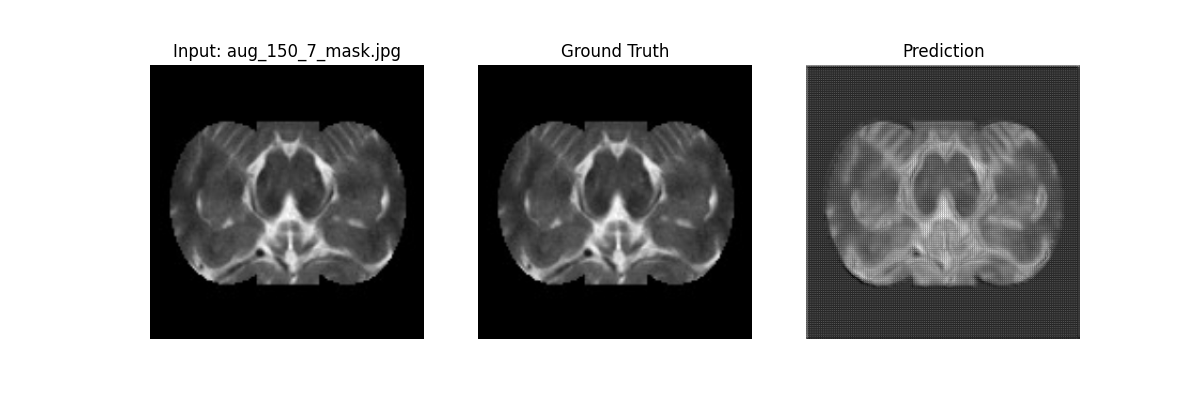
Figure 1
References: 1. Mahapatra C., Manchanda R. Computational assessment of calcium channel effects on subthalamic nucleus neuronal cells: abnormal bursting patterns in Parkinson’s disease. Movement Disorders. 2016.
2. Mei J. et al. Machine learning for the diagnosis of Parkinson’s disease: a review. Frontiers in Aging Neuroscience. 2021.
To cite this abstract in AMA style:
G. Kuanar, C. Mahapatra. Automated MRI Biomarker Extraction for Parkinson’s Disease Using Attention-Based U-Net Segmentation [abstract]. Mov Disord. 2026; 41 (suppl 1). https://www.mdsabstracts.org/abstract/automated-mri-biomarker-extraction-for-parkinsons-disease-using-attention-based-u-net-segmentation/. Accessed October 1, 2026.« Back to 2026 International Congress
MDS Abstracts - https://www.mdsabstracts.org/abstract/automated-mri-biomarker-extraction-for-parkinsons-disease-using-attention-based-u-net-segmentation/