
Objective: To investigate clinical gait characteristics and cerebellar-pedunculopontine nucleus (PPN) functional connectivity (FC) changes in early-stage and moderate-to-advanced-stage Parkinson’s disease (PD) with gait disturbance using wearable sensing technology and resting-state functional magnetic resonance imaging (rs‑fMRI), and clarify the cerebellar-PPN circuit’s role in gait dysfunction progression.
Background: Gait disturbance is a core PD motor symptom, increasing fall risk and impairing quality of life [1,2]. The cerebellum and PPN regulate motor control and balance, and their abnormal FC may be linked to PD gait disturbance onset and progression [3,4], but its stage-specific alterations remain unclear.
Method: A total of 40 PD patients with gait disturbance (22 early-stage, 18 moderate-to-advanced-stage) were enrolled from the outpatient and inpatient departments of Zhongnan Hospital of Wuhan University, Wuhan, China. The study received ethical approval from the Medical Ethical Committee of Zhongnan Hospital of Wuhan University. Demographics (sex, age, BMI) and disease duration were collected. Gait parameters were quantified using wearable sensing technology, including gait variability, bilateral stride length, walking speed, swing/stance phase duration, and forefoot/heel pressure. Cerebellar-PPN resting-state FC (rs-FC) was measured via rs-fMRI, with between-group comparisons using independent samples t-tests.
Results: No significant between‑group differences in age, sex, BMI or disease duration (all p>0.05). Compared with the early‑stage group, the moderate‑to‑advanced‑stage group showed markedly increased gait variability (p=0.0041), reduced bilateral stride length (p=0.0026) and walking speed (p<0.0001), shortened swing phase, prolonged stance phase (all p<0.01), and highly significant abnormalities in plantar anterior–posterior pressure distribution (p<0.0001) [table 1]. Rs‑fMRI showed significantly higher cerebellar-PPN rs‑FC Z‑value in the moderate‑to‑advanced‑stage group (p<0.001) [figure 1].
Conclusion: PD gait disturbance progression is accompanied by dynamic balance and propulsion impairment, together with pathologically or compensatory increased cerebellar-PPN FC. The cerebellar-PPN circuit is a key pathophysiological component of PD gait disturbance, providing a potential neural circuit target for precise intervention.
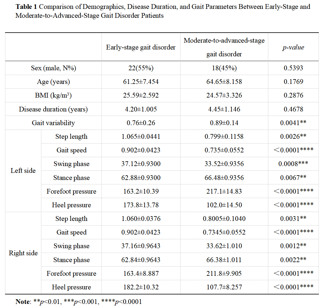
Table 1
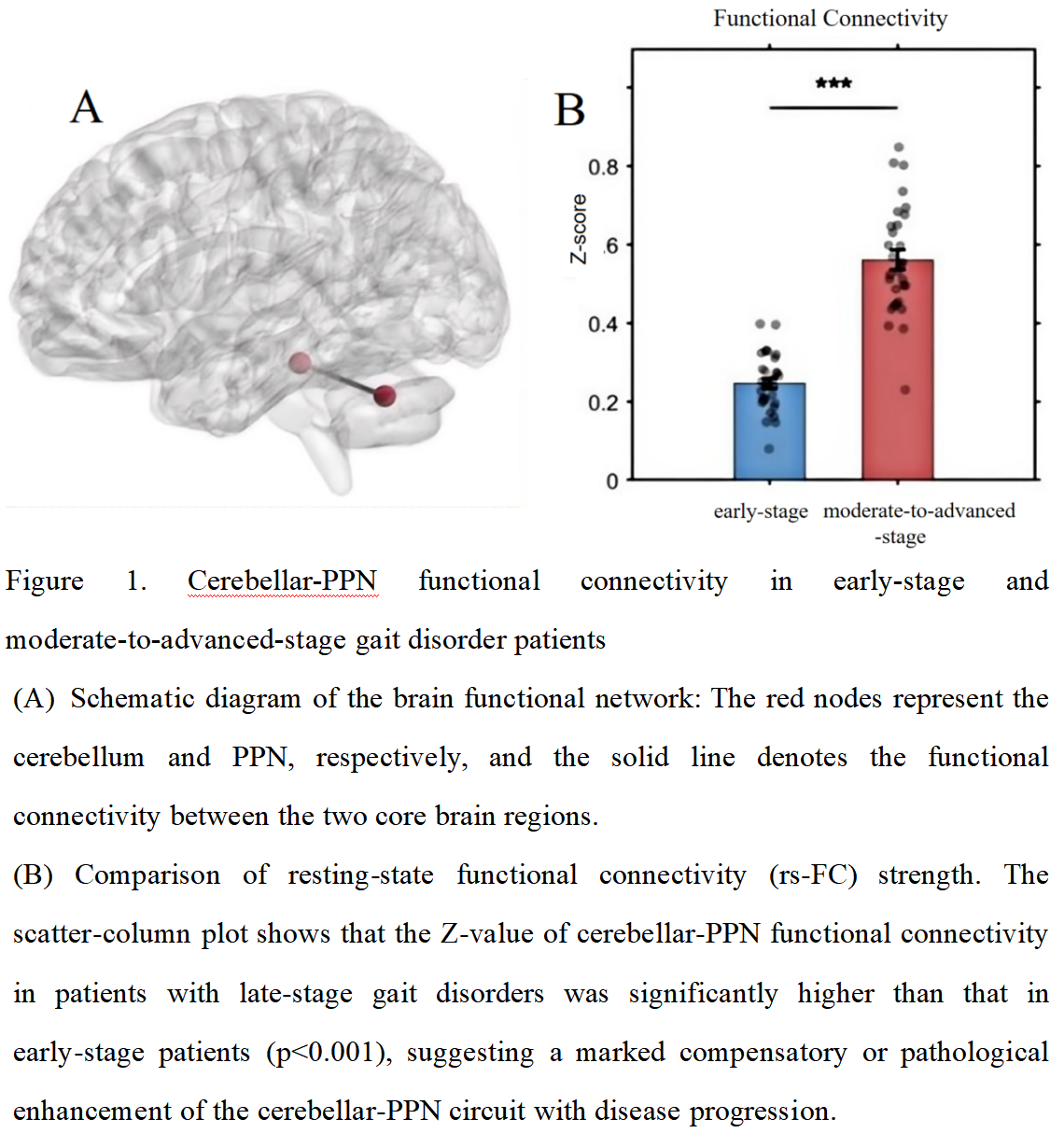
Figure 1
References: [1] DE PASQUALE P, BONANNO M, DE MARCHIS C, et al. Beyond clinical scales: an observational study on instrumental gait analysis and biomechanical patterns in patients with Parkinson’s disease [J]. Front Bioeng Biotechnol, 2025, 13: 1541240.
[2] GUO Y, YANG J, LIU Y, et al. Detection and assessment of Parkinson’s disease based on gait analysis: A survey [J]. Front Aging Neurosci, 2022, 14: 916971.
[3] FRENCH I T, MUTHUSAMY K A. A Review of the Pedunculopontine Nucleus in Parkinson’s Disease [J]. Front Aging Neurosci, 2018, 10: 99.
[4] MAITI B, RAWSON K S, TANENBAUM A B, et al. Functional Connectivity of Vermis Correlates with Future Gait Impairments in Parkinson’s Disease [J]. Mov Disord, 2021, 36(11): 2559-68.
To cite this abstract in AMA style:
J. Su, D. Liu, J. Shang, L. She, H. Wang, Y. Pan. Clinical Gait Traits and Cerebellar-PPN Functional Connectivity Abnormalities in Early and Moderate-to-Advanced PD with Gait Disturbance via Perceptual Technology and MRI [abstract]. Mov Disord. 2026; 41 (suppl 1). https://www.mdsabstracts.org/abstract/clinical-gait-traits-and-cerebellar-ppn-functional-connectivity-abnormalities-in-early-and-moderate-to-advanced-pd-with-gait-disturbance-via-perceptual-technology-and-mri/. Accessed October 1, 2026.« Back to 2026 International Congress
MDS Abstracts - https://www.mdsabstracts.org/abstract/clinical-gait-traits-and-cerebellar-ppn-functional-connectivity-abnormalities-in-early-and-moderate-to-advanced-pd-with-gait-disturbance-via-perceptual-technology-and-mri/