
Category: Parkinson's disease: Neuroimaging
Objective: To determine whether cerebellar axonal microstructural abnormality is associated with cognitive performance in PD.
Background: Cognitive impairment in Parkinson’s disease (PD) often affects executive function, attention, visuospatial skills, and processing speed [1]. While frontostriatal dysfunction is well established [2], neuroimaging suggests altered cerebellar structure and connectivity in PD, which may contribute to cognitive deficits [3, 4]. The role of cerebellar microstructure in domain-specific cognition, however, remains unclear and may offer insights into the neural mechanisms of cognitive impairment in PD.
Method: We studied 154 patients with PD from the Parkinson’s Progression Markers Initiative (PPMI) [5]. Using diffusion MRI data, quantitative anisotropy (QA) values, representing axonal density, were extracted from cerebellar regions using the SUIT atlas [6]. A controls-referenced Mahalanobis distance metric (MD_QA) was computed to quantify global cerebellar microstructural abnormality, with higher values indicating a greater reduction in axonal density compared to healthy controls [7]. Associations between MD_QA and cognitive performance—global cognition (Montreal Cognitive Assessment [MoCA]), memory (Hopkins Verbal Learning Test–Revised [HVLT-R; immediate and delayed recall]), visuospatial function (Benton Judgment of Line Orientation [JLO]), processing speed (Symbol Digit Modalities Test [SDMT]), and working memory (Letter-Number Sequencing [LNS]) and language (semantic fluency [SF])—were examined using linear regression models adjusted for age and sex. False discovery rate (FDR; q-values) correction was applied across cognitive outcomes.
Results: Higher MD_QA was significantly associated with lower SDMT performance (β = −1.30, q = 0.008), lower SF (β = −1.36, q = 0.01), and lower HVLT-R (β = −0.65, q = 0.01). Other cognitive measures were not significantly associated with MD_QA after correction (Figure 1).
Conclusion: Increased cerebellar microstructural abnormality was associated with reduced processing speed, semantic fluency, and verbal learning performance in PD. These findings suggest a relationship between cerebellar integrity and domain-specific cognitive performance in PD. Future analyses will examine regional/lateralized specificity for cognitive associations.
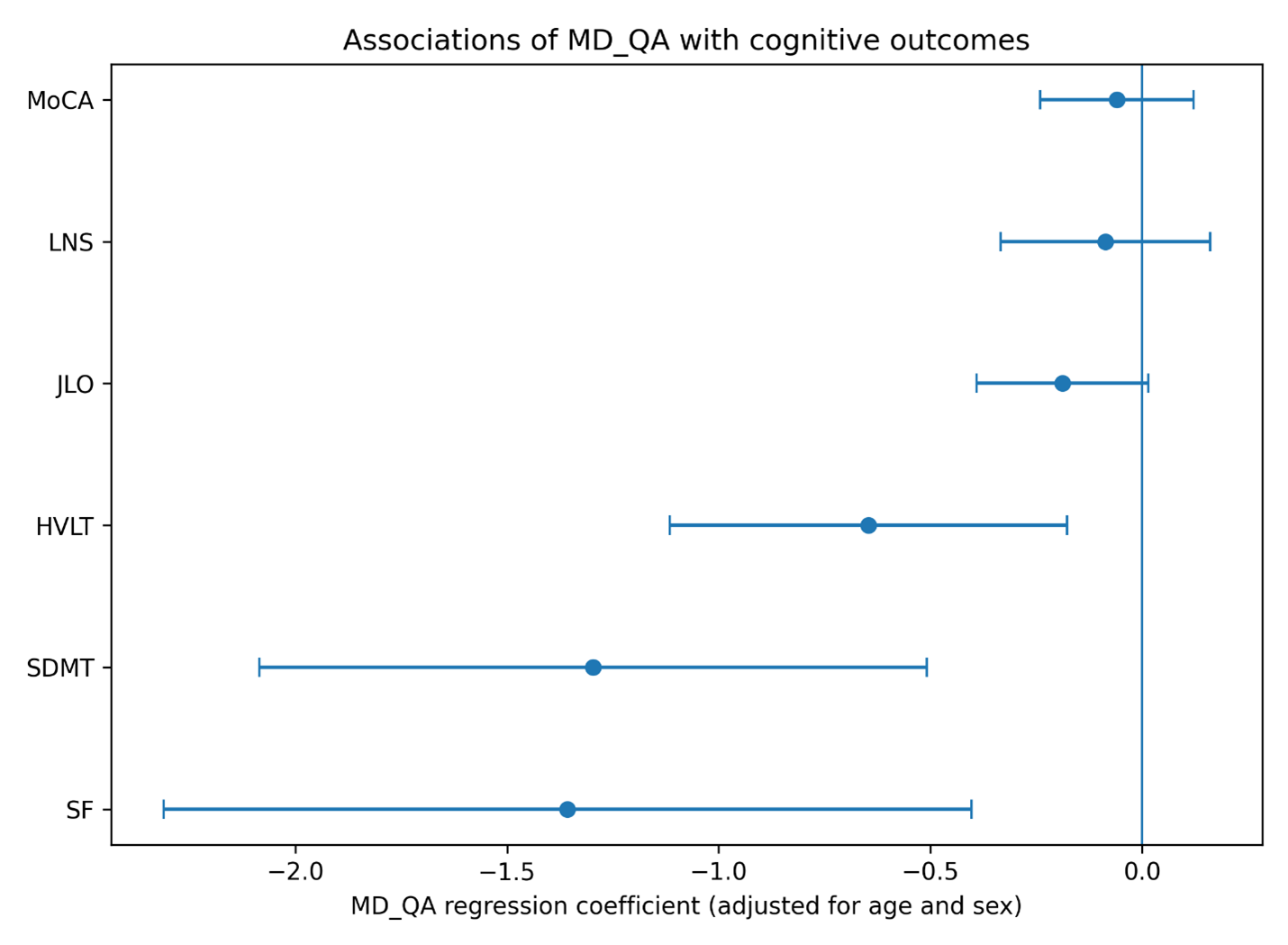
MD QA Associations with Cognition
References: 1. Williams-Gray CH, Foltynie T, Brayne CE, Robbins TW, Barker RA (2007) Evolution of cognitive dysfunction in an incident Parkinson’s disease cohort. Brain 130:1787-98. https://doi.org/10.1093/brain/awm111
2. de la Fuente-Fernández R (2012) Frontostriatal cognitive staging in Parkinson’s disease. Parkinsons Dis 2012:561046. https://doi.org/10.1155/2012/561046
3. Dan X, et al. (2021) Altered Cerebellar Resting-State Functional Connectivity in Early-Stage Parkinson’s Disease Patients With Cognitive Impairment. Front Neurol 12:678013. https://doi.org/10.3389/fneur.2021.678013
4. Bosch TJ, et al. (2021) Altered Cerebellar Oscillations in Parkinson’s Disease Patients during Cognitive and Motor Tasks. Neuroscience 475:185-196. https://doi.org/10.1016/j.neuroscience.2021.08.021
5. Marek K, et al. (2018) The Parkinson’s progression markers initiative (PPMI) – establishing a PD biomarker cohort. Ann Clin Transl Neurol 5:1460-1477. https://doi.org/10.1002/acn3.644
6. Diedrichsen J (2006) A spatially unbiased atlas template of the human cerebellum. NeuroImage 33:127-138. https://doi.org/https://doi.org/10.1016/j.neuroimage.2006.05.056
7. Vijayakumari AA, Mandava N, Hogue O, Fernandez HH, Walter BL (2023) A novel MRI-based volumetric index for monitoring the motor symptoms in Parkinson’s disease. J Neurol Sci 453:120813. https://doi.org/10.1016/j.jns.2023.120813
To cite this abstract in AMA style:
A. Vijayakumari, L. Saadatpour, D. Floden, K. Sakaie, H. Kaur, H. Fernandez, B. Walter. Diffusion MRI Evidence of Cerebellar Involvement in Cognitive Dysfunction in Parkinson’s Disease [abstract]. Mov Disord. 2026; 41 (suppl 1). https://www.mdsabstracts.org/abstract/diffusion-mri-evidence-of-cerebellar-involvement-in-cognitive-dysfunction-in-parkinsons-disease/. Accessed October 1, 2026.« Back to 2026 International Congress
MDS Abstracts - https://www.mdsabstracts.org/abstract/diffusion-mri-evidence-of-cerebellar-involvement-in-cognitive-dysfunction-in-parkinsons-disease/