
Objective: To define the distinct multimodal biomarker profile of Parkinson’s disease with rapid eye movement sleep behavior disorder (PD-RBD) by integrating clinical data, Olink-based plasma proteomics, structural and functional neuroimaging, and to develop an accurate diagnostic model for this subtype.
Background: PD-RBD is an aggressive subtype marked by rapid progression. Its pathophysiology, particularly linking systemic inflammation to brain alterations, is unclear.
Method: This prospective study enrolled 176 participants (45 PD-RBD, 89 PD-nRBD, 42 controls). We assessed 92 inflammatory proteins (Olink), diffusion tensor imaging (DTI) for microstructure and glymphatic function (ALPS index), and resting-state fMRI for interhemispheric connectivity (VMHC). Ensemble machine learning was employed for feature selection and model construction, and mediation analysis was conducted to examine mechanistic relationships.
Results: Proteomic analysis identified significant upregulation of GDNF and downregulation of FGF‑21 in PD-RBD [Figure 1]. Neuroimaging revealed decreased fractional anisotropy in the left supplementary motor area, increased VMHC in the cerebellum and supplementary motor area (SMA), and a reduced ALPS index in PD-RBD [Figure 2]. Ten key multimodal features were selected, and a logistic regression model achieved exceptional diagnostic accuracy (AUC = 0.982) in distinguishing PD-RBD [Figure 3]. Mediation analysis revealed that the effects of FGF21, GDNF, and MCP-4 on PD-RBD were fully mediated by imaging markers: VMHC (for FGF21 and GDNF), ALPS index (for GDNF), and fractional anisotropy in the left SMA (for MCP-4) [Figure 4].
Conclusion: The integration of Olink proteomics with multimodal MRI through machine learning yields a highly accurate biomarker model, providing a robust framework for identifying this aggressive PD subtype and suggesting a link between systemic inflammation and brain connectivity in its pathophysiology.
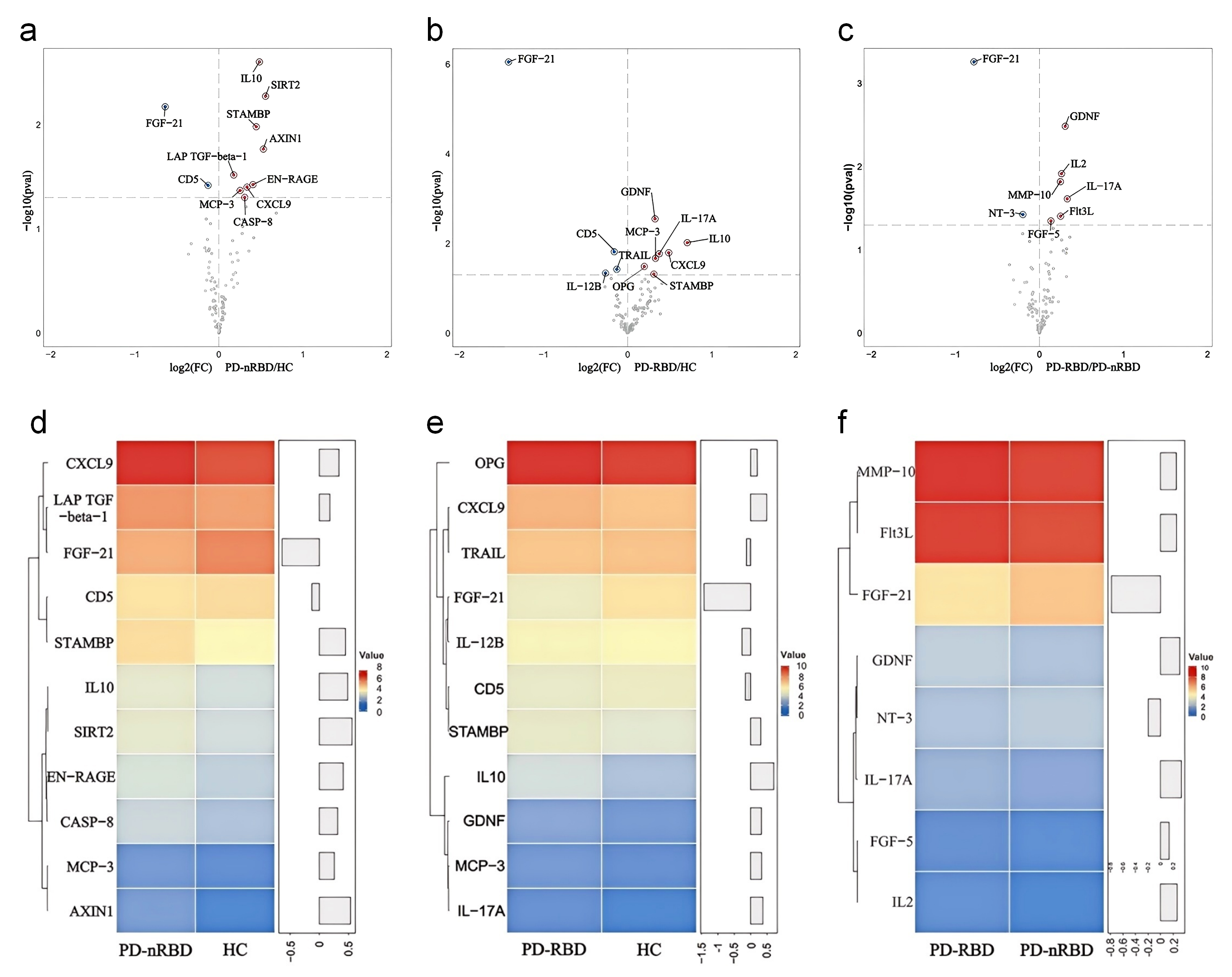
Figure 1. Plasma proteomics in HC, PD-RBD, PD-nRBD
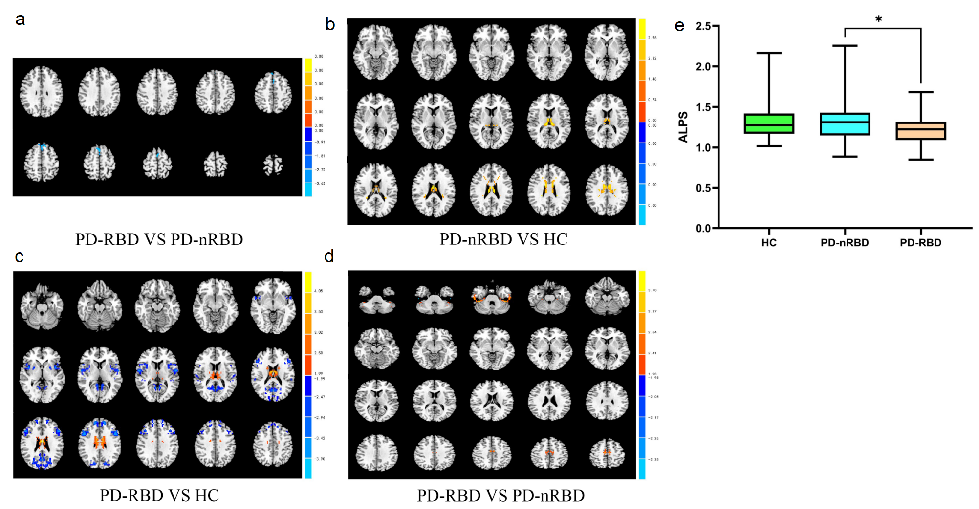
Figure 2. Brain imaging in HC, PD-RBD, PD-nRBD
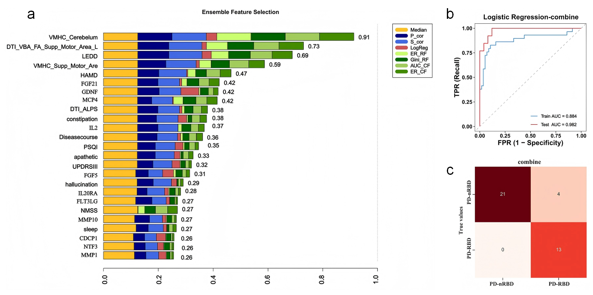
Figure 3. ML model feature & results for PD-RBD
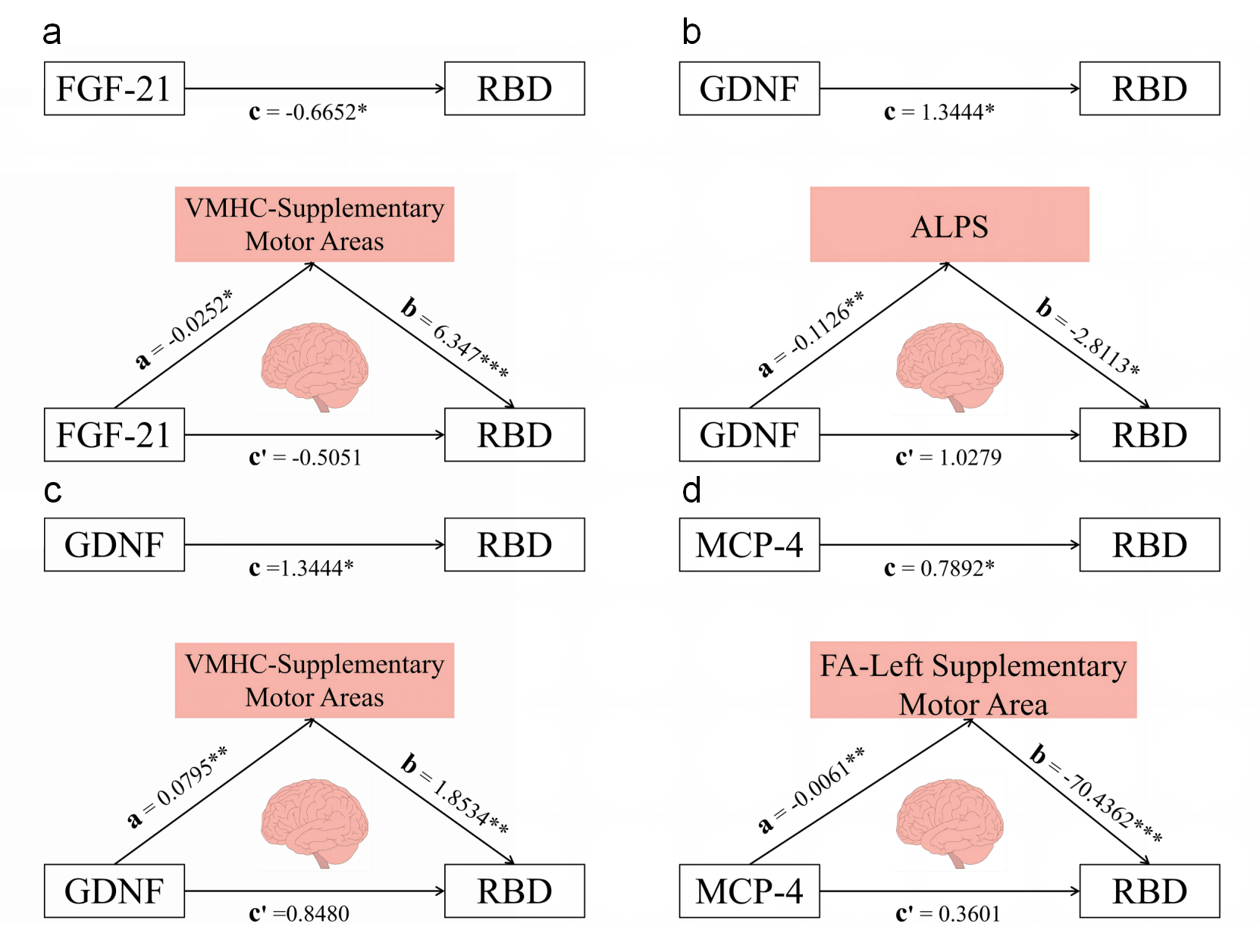
Figure 4. Mediation of imaging & protein in PD-RBD
To cite this abstract in AMA style:
LH. Zhu, F. Wang, YY. Zhu, KF. Yin, CX. Liu, LF. Yang, Q. Cui, WN. Xiong, YB. Han, AL. Pang, XL. Yang. Integrating Olink Proteomics and Multimodal MRI to Reveal a Multimodal Signature of Parkinson’s Disease with REM Sleep Behavior Disorder [abstract]. Mov Disord. 2026; 41 (suppl 1). https://www.mdsabstracts.org/abstract/integrating-olink-proteomics-and-multimodal-mri-to-reveal-a-multimodal-signature-of-parkinsons-disease-with-rem-sleep-behavior-disorder/. Accessed October 1, 2026.« Back to 2026 International Congress
MDS Abstracts - https://www.mdsabstracts.org/abstract/integrating-olink-proteomics-and-multimodal-mri-to-reveal-a-multimodal-signature-of-parkinsons-disease-with-rem-sleep-behavior-disorder/