
Category: MSA, PSP, CBS: Neuroimaging
Objective: To study cortical microstructural alterations in patients with progressive supranuclear palsy (PwPSP) and their associations with both cognitive and clinical symptoms.
Background: Midbrain’s atrophy on structural imaging is widely demonstrated in PwPSP [1]. However, cortical changes are poorly studied. Cortical mean diffusivity (cMD) can be calculated through surface-based diffusion magnetic resonance imaging (MRI) approach to detect microstructural cortical changes. This measure has been suggested as a sensitive marker to detect early degeneration in neurodegenerative diseases [2]. Nevertheless, no previous study has examined microstructural cortical changes through cMD and investigated its association with cognitive and disease severity.
Method: Nineteen PSP and 23 controls ¡ underwent 3D T1- and DWI-weighted magnetic resonance imaging (MRI) on a 3T scanner. Clinical data was collected for patients with PSP, and both groups underwent Mini-mental state examination (MMSE) as a measure of global cognition. Differences in cMD between-groups were explored adjusting for age, sex and years of education, and related to clinical symptoms and MMSE scores. A vertex-wise threshold of p<0.01 was applied [3].
Results: Patients with PSP presented lower years of education and MMSE scores [Table 1]. Patients with PSP showed increased cMD in clusters with peak at the left pars opercularis, superior frontal, parietal and temporal, posterior cingulate and parahippocampal, as well as, the right superior frontal, insula, inferior parietal, and fusiform, with p<0.01. [Figure 1]. Progressive Supranuclear Palsy Rating Scale (PSPRS) scores correlated positively with cMD in the left isthmus of the cingulate gyrus and superior frontal, and the right pars triangularis [Figure 2]. Moreover, a negative correlation was found between MMSE scores and the right inferior temporal [Figure 2].
Conclusion: cMD reveals widespread microstructural changes suggesting cortical degeneration across bilateral frontal, parietal, and temporal regions in PSP. Notably, higher cMD values are associated with greater disease severity and poorer global cognitive performance, highlighting its potential as an imaging biomarker for monitoring cortical neurodegeneration and tracking disease progression.
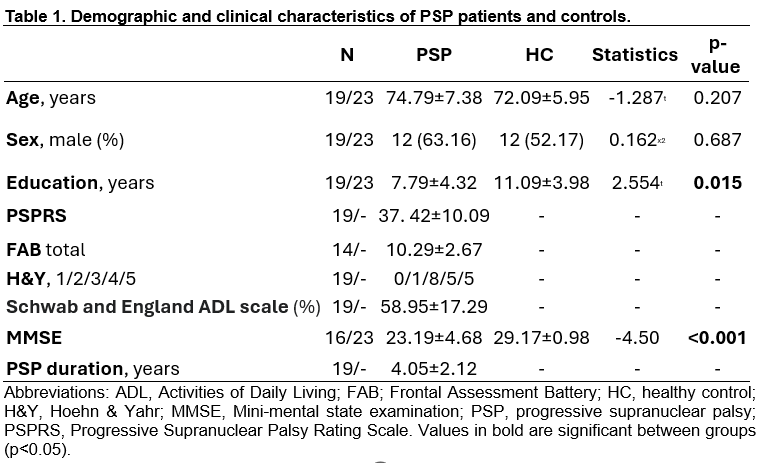
Table 1.
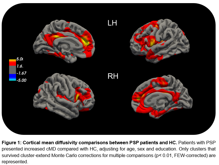
Figure 1.
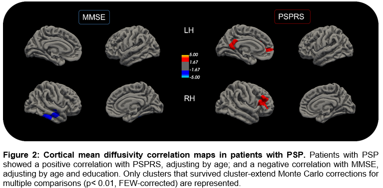
Figure 2.
References: [1] Keir G, Roytman M, Mashriqi F, Shahsavarani S, Franceschi AM. Atypical Parkinsonian Syndromes: Structural, Functional, and Molecular Imaging Features. AJNR Am J Neuroradiol. 2024 Dec 9;45(12):1865-1877. doi: 10.3174/ajnr.A8313.
[2] Sun, P., He, Z., Li, A., Yang, J., Zhu, Y., Cai, Y., Ma, T., Ma, S., Guo, T., & Alzheimer’s Disease Neuroimaging Initiative (2024). Spatial and temporal patterns of cortical mean diffusivity in Alzheimer’s disease and suspected non-Alzheimer’s disease pathophysiology. Alzheimer’s & dementia: the journal of the Alzheimer’s Association, 20(10), 7048–7061. https://doi.org/10.1002/alz.14176
[3] Montal V, Vilaplana E, Alcolea D, Pegueroles J, Pasternak O, González-Ortiz S, et al. Cortical microstructural changes along the Alzheimer’s disease continuum. Alzheimer’s and Dementia. 2018 Mar 1;14(3):340–51.
To cite this abstract in AMA style:
C. Garcia Vicente, J. Pardo, C. Martín-Barceló, I. Roura, C. Falcon, R. Sala-Llonch, A. Campabadal, N. Bargalló, O. Brengaret, C. Brenlla, C. Painous, A. Cámara, M. Martí, Y. Compta, C. Junqué, B. Segura. Microstructural cortical alterations are related to cognitive and disease severity in progressive supranuclear palsy [abstract]. Mov Disord. 2026; 41 (suppl 1). https://www.mdsabstracts.org/abstract/microstructural-cortical-alterations-are-related-to-cognitive-and-disease-severity-in-progressive-supranuclear-palsy/. Accessed October 1, 2026.« Back to 2026 International Congress
MDS Abstracts - https://www.mdsabstracts.org/abstract/microstructural-cortical-alterations-are-related-to-cognitive-and-disease-severity-in-progressive-supranuclear-palsy/