
Objective: To characterize patterns of regional glucose hypometabolism on Fluorodeoxyglucose (FDG)-PET and structural atrophy on MRI across clinical variants of progressive supranuclear palsy (PSP).
Background: PSP encompasses a spectrum of clinical variants beyond the classical Richardson syndrome (PSP-RS), including cortical and subcortical variants. Neuroimaging biomarkers that capture this clinical heterogeneity remain incompletely defined.
Method: Seventeen patients with PSP were prospectively evaluated regarding demographics, clinical profile, and clinical variants in the first 3 years and current clinical variant at baseline. All underwent brain FDG-PET and MRI on a hybrid PET/MRI scan. Quantitative group analyses with SPM12 were performed and compared with 17 age-matched healthy controls using voxel-based analysis. MRI data were analyzed using SPM-based voxel-based morphometry. For subgroup analyses, patients were grouped into cortical, RS/classical, and subcortical phenotypic clusters.
Results: PSP patients had a mean age of 69.9 ± 8.3 years, were 64.7% male, and had a disease duration of 4.8 ± 2.3 years. Clinical variants during the first three years were diverse (PSP-RS=6, PSP-PI=2, PSP-OM=1, PSP-P=4, PSP-PGF=3, PSP-CBS=1, PSP-SL=1), with 94% converging to probable PSP-RS at final consensus review. Whole-group MRI volumetry revealed bilateral basal ganglia atrophy, while FDG-PET showed hypometabolism in the midbrain peduncle, medial frontal cortex, inferior frontal cortex, and basal ganglia [Figures 1, 2]. Subgroup analyses showed distinct metabolic signatures: PSP-RS (n=6) exhibited dorsal midbrain hypometabolism [Figure 3]; cortical variants (n=2) showed hypometabolism in the left supplementary motor and medial frontal cortices [Figure 4]; and subcortical variants (n=9) demonstrated diffuse midbrain hypometabolism, most prominent in the midbrain peduncle [Figure 5].
Conclusion: FDG-PET depicts PSP-specific metabolic patterns among clinical variants. Midbrain hypometabolism was a consistent feature across groups, while frontal and SMA involvement distinguished cortical phenotypes. These results support the utility of FDG-PET in characterizing the clinical heterogeneity of PSP and may inform future biomarker studies.
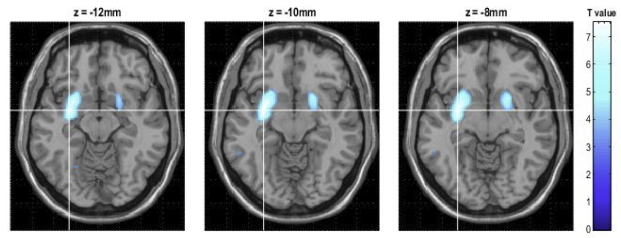
VBM: bilateral basal ganglia atrophy (PSP < HC)
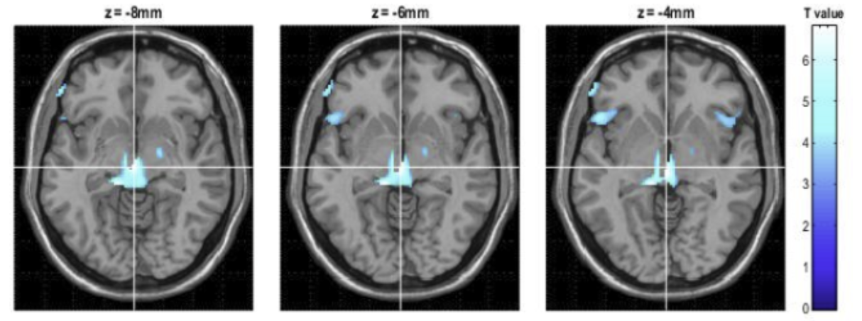
FDG-PET: whole-group hypometabolism (PSP < HC)
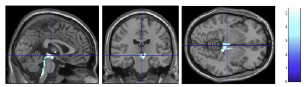
FDG-PET: dorsal midbrain hypometabolism (PSP-RS)
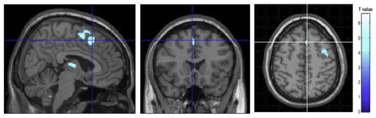
FDG-PET: frontal hypometabolism, cortical PSP
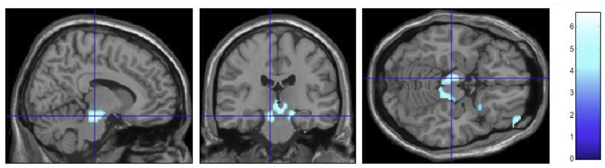
FDG-PET: midbrain hypometabolism, subcortical PSP
References: Martí-Andrés G, van Bommel L, Meles SK, Riverol M, Valentí R, Kogan RV, Renken RJ, Gurvits V, van Laar T, Pagani M, Prieto E, Luquin MR, Leenders KL, Arbizu J. Multicenter Validation of Metabolic Abnormalities Related to PSP According to the MDS-PSP Criteria. Mov Disord. 2020 Nov;35(11):2009-2018.
Ali F, Clark H, Machulda M, Senjem ML, Lowe VJ, Jack CR Jr, Josephs KA, Whitwell J, Botha H. Patterns of brain volume and metabolism predict clinical features in the progressive supranuclear palsy spectrum. Brain Commun. 2024 Jul 16;6(4):fcae233
Wang H, Wang B, Liao Y, Niu J, Chen M, Chen X, Dou X, Yu C, Zhong Y, Wang J, Jin N, Kang Y, Zhang H, Tian M, Luo W. Identification of metabolic progression and subtypes in progressive supranuclear palsy by PET molecular imaging. Eur J Nucl Med Mol Imaging. 2025 Feb;52(3):823-835.
To cite this abstract in AMA style:
V. Maciel, M. de Oliveira, G. Martins, C. Carneiro, P. Barbosa, C. Buchpiguel, E. Barbosa, A. Coutinho, J. Parmera. Metabolic-Structural Patterns Across Progressive Supranuclear Palsy Clinical Variants: a Multimodal PET-MRI Study [abstract]. Mov Disord. 2026; 41 (suppl 1). https://www.mdsabstracts.org/abstract/metabolic-structural-patterns-across-progressive-supranuclear-palsy-clinical-variants-a-multimodal-pet-mri-study/. Accessed October 1, 2026.« Back to 2026 International Congress
MDS Abstracts - https://www.mdsabstracts.org/abstract/metabolic-structural-patterns-across-progressive-supranuclear-palsy-clinical-variants-a-multimodal-pet-mri-study/