
Objective: This study aims to clarify subtype-specific changes in mesencephalic locomotor region (MLR)-cerebellar functional connectivity(FC)[1][2] between body-first and brain-first Parkinson’s disease (PD) patients[3][4], and to further characterize the correlation between these changes and gait disturbance. By identifying unique connectivity patterns, and the targeting of personalized temporal interference stimulation (TIS)[5][6].
Background: PD can be classified into body-first and brain-first subtypes based on rapid eye movement sleep behavior disorder(RBD), with distinct clinical manifestations. Gait disturbance is a core motor symptom of PD, and the MLR-cerebellar network is critical for gait regulation. However, differences in FC between these two subtypes and their association with gait impairment remain unclear.
Method: Thirty brain-first and 30 body-first participants were recruited via purposeful sampling, with baseline data and phenotypic classification completed (Table 1). Using the MLR and Pedunculopontine Nucleus(PPN) as seed regions, we analyzed multi-brain region functional connectivity. Mean FC values of regions with significant differences were extracted for statistics. Pearson correlation was performed between MLR-cerebellar FC and gait variability(GV).
Results: In body-first PD patients, MLR-seeded FC revealed significantly enhanced connectivity with the brainstem/cerebellum and reduced connectivity with the primary motor cortex (M1) and supplementary motor area (SMA) (Fig. 1A). In brain-first PD patients, no MLR-specific abnormalities were observed (Fig. 1B). Voxel-wise whole-brain analysis with the PPN as seed showed that body-first PD exhibited significantly increased connectivity in the bilateral posterior cerebellar lobes and cerebellar vermis compared to brain-first PD (Fig. 2). Quantitative comparison demonstrated significantly higher brainstem-cerebellum connectivity (Z-score) in the body-first group than in the brain-first group (Fig. 3). Furthermore, Pearson correlation analysis revealed a significant positive correlation between MLR-cerebellum connectivity and GV in body-first PD (Fig. 4).
Conclusion: Distinct brain network mechanisms by RBD subtype were identified, with MLR-cerebellar connectivity positively correlated with GV in body-first patients, informing predictive models and targeted TIS therapies.
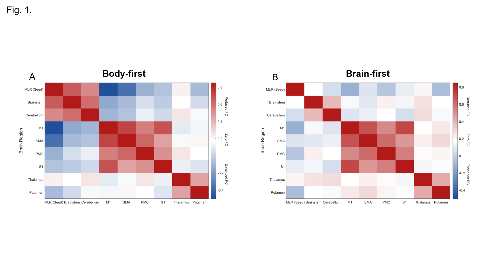
Fig.1 Functional connectivity matrix plot
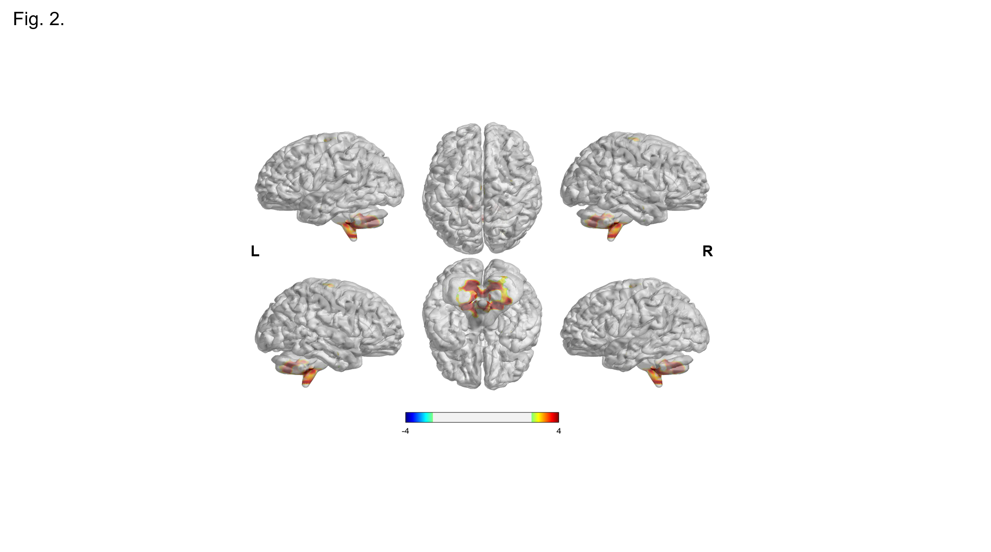
Fig.2 functional connectivity analysis.
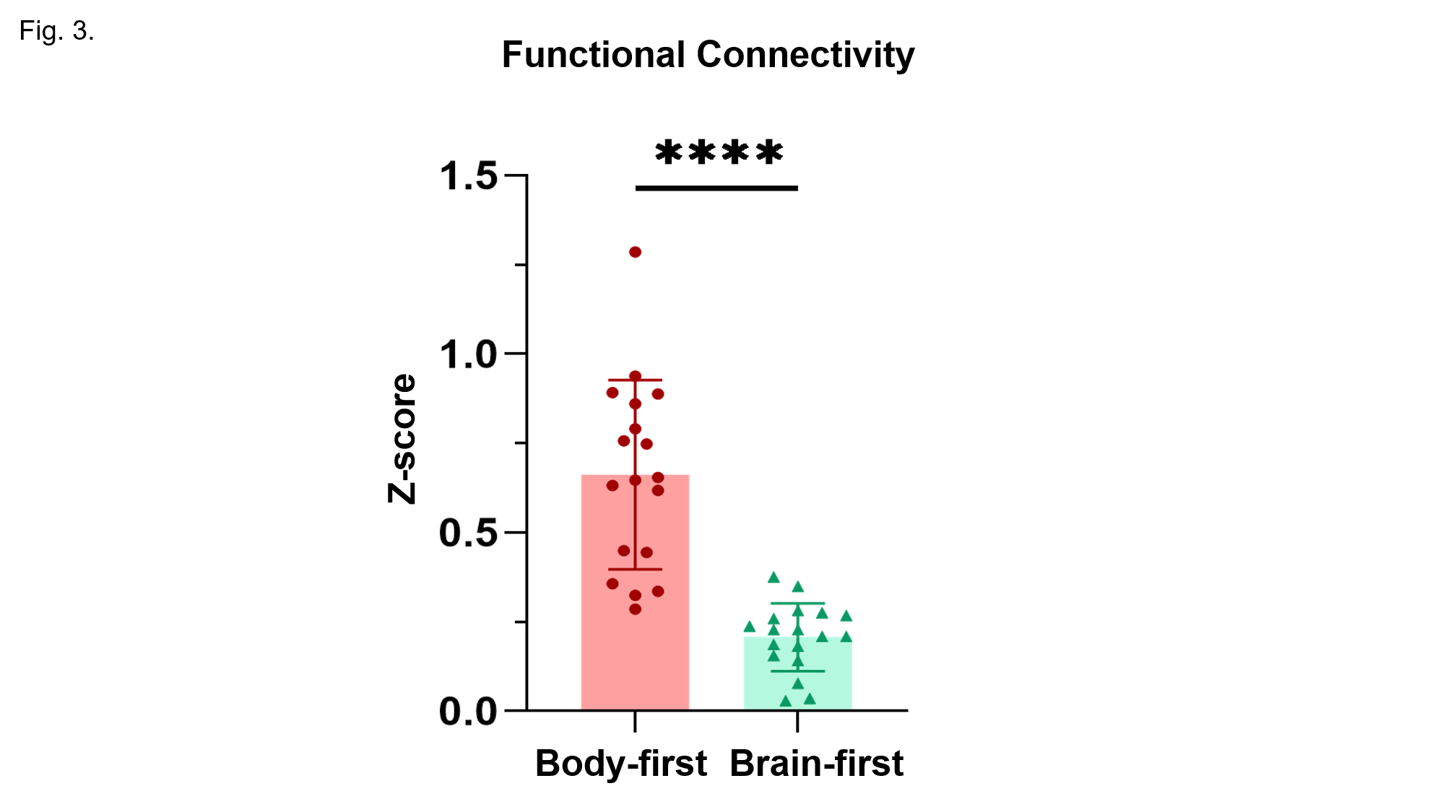
Fig.3 functional connectivity strength.
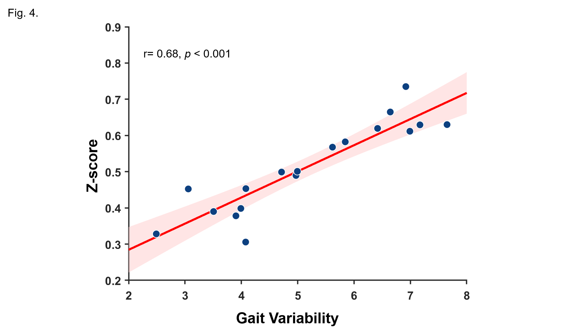
Fig.4 FC strength and gait variability.
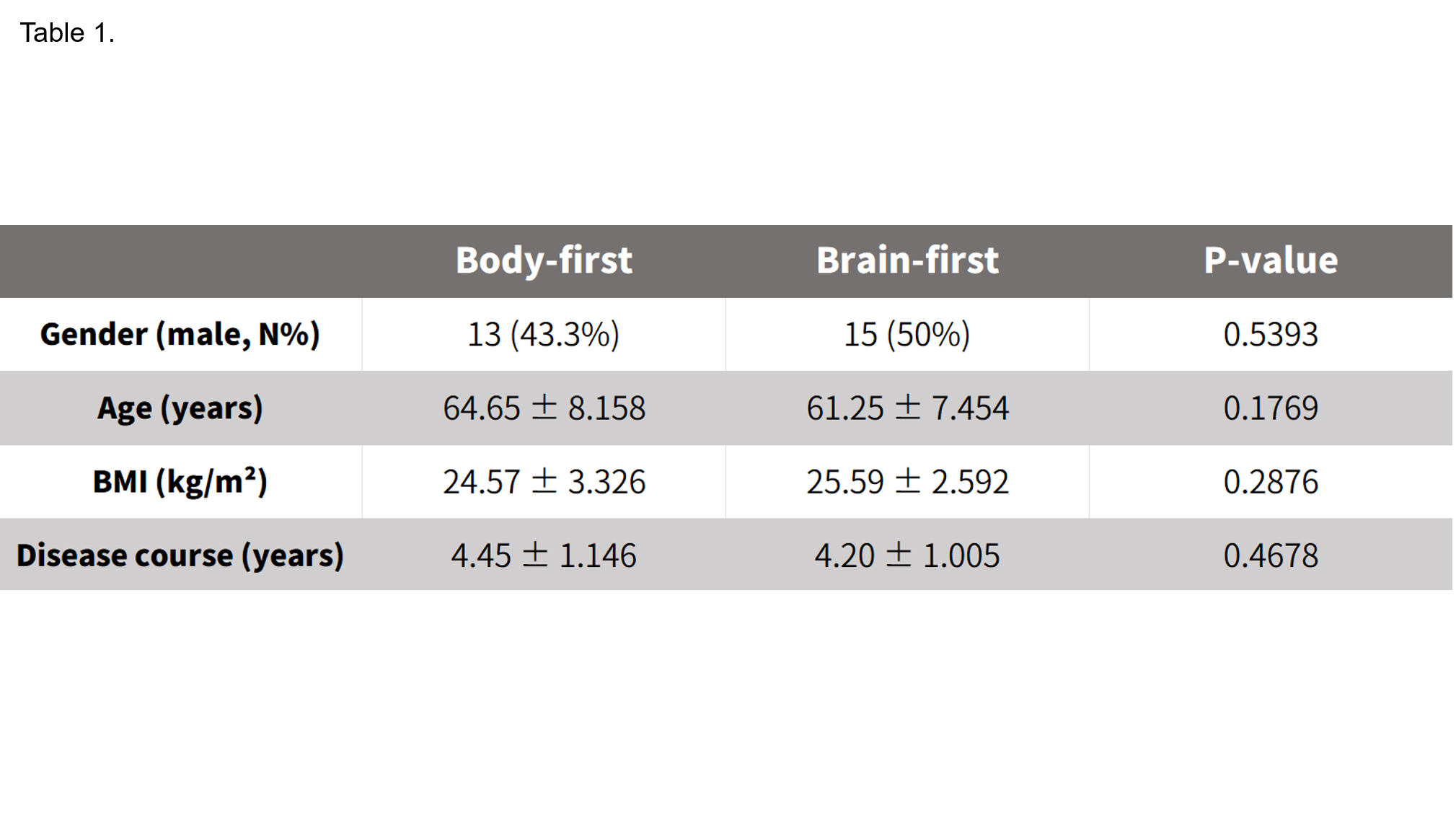
Table.1 Table of Participant Composition
References: [1]Ferreira-Pinto MJ, Kanodia H, Falasconi A, Sigrist M, Esposito MS, Arber S. Functional diversity for body actions in the mesencephalic locomotor region. Cell. 2021 Aug 19;184(17):4564-4578.e18. doi: 10.1016/j.cell.2021.07.002. Epub 2021 Jul 23. PMID: 34302739; PMCID: PMC8382160
[2]Wang S, Xiao Y, Hou Y, Li C, Lin J, Yang T, Che N, Jiang Q, Zheng X, Liu J, Shang H. Altered gait speed and brain network connectivity in Parkinson’s disease. Cereb Cortex. 2024 Nov 5;34(11):bhae429. doi: 10.1093/cercor/bhae429. PMID: 39505570.
[3]Horsager J, Knudsen K, Sommerauer M. Clinical and imaging evidence of brain-first and body-first Parkinson’s disease. Neurobiol Dis. 2022 Mar;164:105626. doi: 10.1016/j.nbd.2022.105626. Epub 2022 Jan 11. PMID: 35031485.
[4]Horsager J, Borghammer P. Brain-first vs. body-first Parkinson’s disease: An update on recent evidence. Parkinsonism Relat Disord. 2024 May;122:106101. doi: 10.1016/j.parkreldis.2024.106101. Epub 2024 Mar 15. PMID: 38519273.
[5]Qi S, Yu J, Li L, Dong C, Ji Z, Cao L, Wei Z, Liang Z. Advances in non-invasive brain stimulation: enhancing sports performance function and insights into exercise science. Front Hum Neurosci. 2024 Nov 29;18:1477111. doi: 10.3389/fnhum.2024.1477111. PMID: 39677404; PMCID: PMC11638246.
[6]Xu S, Cui H, Xiao X, Manshaii F, Hong G, Chen J. Precision at Deep Brain: Noninvasive Temporal Interference Stimulation. ACS Nano. 2025 Nov 25;19(46):39589-39614. doi: 10.1021/acsnano.5c15238. Epub 2025 Nov 13. PMID: 41232023.
To cite this abstract in AMA style:
J. Jin, Z. Zong, S. Si, S. She, W. Wang, P. Pan. MLR–cerebellar network in body-first vs. brain-first PD: functional connectivity and clinical correlation with gait impairment [abstract]. Mov Disord. 2026; 41 (suppl 1). https://www.mdsabstracts.org/abstract/mlr-cerebellar-network-in-body-first-vs-brain-first-pd-functional-connectivity-and-clinical-correlation-with-gait-impairment/. Accessed October 1, 2026.« Back to 2026 International Congress
MDS Abstracts - https://www.mdsabstracts.org/abstract/mlr-cerebellar-network-in-body-first-vs-brain-first-pd-functional-connectivity-and-clinical-correlation-with-gait-impairment/