
Category: Parkinson's disease: Neuroimaging
Objective: To describe the autonomic function profile and quantitative brainstem substructures analysis in a 62-year-old male of Parkinsonism complicated by severe autonomic failure and neuropsychiatric symptoms.
Background: A 62-year-old male presented with a six-year history of asymmetric Parkinsonism characterized by left-sided resting tremor, bradykinesia, rigidity, and a shuffling gait. Motor symptoms stabilized following the introduction of levodopa/benserazide and entacapone. The clinical course was subsequently complicated by weekly episodes of postprandial paroxysmal somnolence and significant orthostatic hypotension. Furthermore, the patient developed “on-state” behavioral disturbances characterized by purposeless, stereotyped actions (e.g., rhythmic table-drumming with various objects) and singing without awareness.
Method: Autonomic Function Testing evaluated both parasympathetic and sympathetic integrity. Parasympathetic function was assessed via the 30:15 ratio, deep breathing heart rate variability and the Valsalva maneuver. Sympathetic response was measured through orthostatic vitals (ΔBP/ΔHR) and peripheral tactile stimulation. T1-weighted MRI data were processed using FreeSurfer (v.7.4.1). Subcortical volumes were extracted via volume-based stream segmentation, and brainstem were analyzed using the Brainstem Substructures Module. (1)
Results: Autonomic Function Testing confirmed global dysautonomia. Parasympathetic failure was evidenced by a blunted 30:15 ratio, reduced E/I ratio during deep breathing, and abnormal Valsalva ratio. Sympathetic integrity was significantly impaired with orthostatic blood pressure drop of 67 mmHg without a compensatory heart rate increase. Preliminary FreeSurfer analysis utilizing the brainstem substructures module is underway to correlate structural atrophy with observed autonomic failure.
Conclusion: We report a case of levodopa-responsive Parkinsonism complicated by profound autonomic failure and stereotyped behaviors. While the clinical presentation mimics MSA, the therapeutic response suggests IPD with severe brainstem involvement. Quantitative analysis of the medullary nuclei via FreeSurfer offers objective evidence of structural damage in the autonomic centers, emphasizing the utility of advanced neuroimaging in characterizing complex Parkinsonian syndromes.
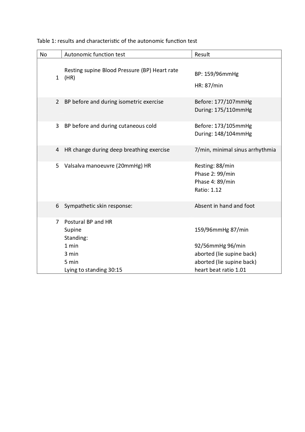
Table 1
References: 1. Iglesias, J.E., Van Leemput, K., Bhatt, P., Casillas, C., Dutt, S., Schuff, N., Truran-Sacrey, D., Boxer, A., and Fischl, B. Bayesian segmentation of brainstem structures in MRI. NeuroImage, 113, June 2015, 184-195.
To cite this abstract in AMA style:
M L. Chin, A K. Lee. Beyond Motor Symptoms: Utilizing the FreeSurfer Brainstem Substructures Module to Characterize a Complex Parkinsonian Phenotype with Profound Dysautonomia [abstract]. Mov Disord. 2026; 41 (suppl 1). https://www.mdsabstracts.org/abstract/beyond-motor-symptoms-utilizing-the-freesurfer-brainstem-substructures-module-to-characterize-a-complex-parkinsonian-phenotype-with-profound-dysautonomia/. Accessed October 1, 2026.« Back to 2026 International Congress
MDS Abstracts - https://www.mdsabstracts.org/abstract/beyond-motor-symptoms-utilizing-the-freesurfer-brainstem-substructures-module-to-characterize-a-complex-parkinsonian-phenotype-with-profound-dysautonomia/