
Objective: To describe the electrophysiological changes that occur in the brain with the use of foscarbidopa-foslevodopa and compare it to other common formulations of levodopa in Parkinson’s disease.
Background: While foscarbidopa-foslevodopa is FDA-approved for subcutaneous Parkinson’s disease (PD) therapy, its impact on brain electrophysiology in patients with deep brain stimulation (DBS) remains unexplored. This study evaluates local field potential recordings (LFP) from DBS-implanted patients to compare the electrophysiological effects of this novel infusion against traditional levodopa formulations.
Method: LFPs were recorded from three PD patients with bilateral sensing-enabled globus pallidus internus DBS systems. Subjects presented in the medication-OFF state and received one of three levodopa formulations: foscarbidopa-foslevodopa (initial loading dose), immediate-release, or extended-release carbidopa-levodopa. Using the BrainSense streaming function with stimulation at 0mA, LFPs were recorded and spectral power was calculated via Welch’s method for theta (4–8 Hz), alpha (8–12 Hz), low beta (13–20 Hz), high beta (21–30 Hz), and gamma (30–100 Hz) bands. Linear regressions assessed significant temporal changes in spectral power as patients transitioned to the medication ON state.
Results: Foscarbidopa-foslevodopa significantly reduced theta and low beta power in both the left (p = 0.0029, p = 0.0037) and right (p = 0.0094, p = 0.0028) hemispheres, with additional alpha reductions in the right (p = 0.0046). In contrast, immediate-release carbidopa-levodopa induced extensive power decreases across alpha, low beta, high beta, and gamma bands in the left hemisphere (all p = 0.0000) and theta (p = 0.0000), low beta (p = 0.0163), high beta (p = 0.0000), and gamma (p = 0.0056) bands in the right. The extended-release formulation yielded significant left-hemisphere reductions in alpha (p = 0.0043), low beta (p = 0.0000), high beta (p = 0.0000), and gamma (p = 0.0221) bands, while the right hemisphere showed decreases across theta (p = 0.0192), alpha (p = 0.0177), and the low beta, high beta, and gamma bands (all p = 0.0000).
Conclusion: We observed all formulations of levodopa modulated low beta, however high beta modulation was limited to carbidopa levodopa immediate release and extended release. The temporal dynamics of levodopa formulations can be insightful for adaptive DBS paradigms that track these frequency bands.
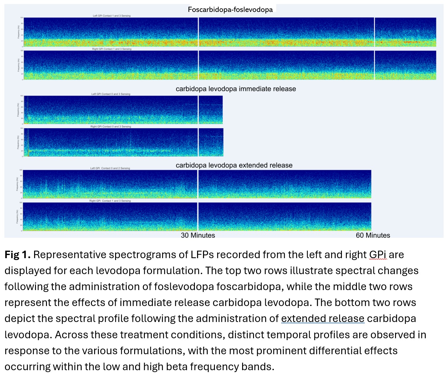
Figure 1 – Levodopa LFP Comparison
To cite this abstract in AMA style:
A. Madamangalam, J. Cagle, C. de Hemptinne, J. Wong. Various Levodopa Formulation effects on Local Field Potentials in Parkinson’s Disease [abstract]. Mov Disord. 2026; 41 (suppl 1). https://www.mdsabstracts.org/abstract/various-levodopa-formulation-effects-on-local-field-potentials-in-parkinsons-disease/. Accessed October 1, 2026.« Back to 2026 International Congress
MDS Abstracts - https://www.mdsabstracts.org/abstract/various-levodopa-formulation-effects-on-local-field-potentials-in-parkinsons-disease/