
Category: MSA, PSP, CBS: Neuroimaging
Objective: To investigate the regional perfusion patterns using PET imaging across various clinical subtypes of PSP and CBD.
Background: On MRI, PSP typically shows midbrain atrophy, whereas CBD is characterized by asymmetric fronto-parietal atrophy. [¹⁸F]FDG PET likewise demonstrate significant hypometabolism in the corresponding regions.
Because a dual-phase [¹⁸F]FP-CIT PET scan provides both a perfusion scan and a DAT scan, we investigated the perfusion patterns obtained from FP-CIT PET in patients with PSP and CBD.
Method: The study included 26 patients with PSP, and 30 patients with CBD. All participants underwent evaluation with the UPDRS Part III and a battery of tests assessing cognitive and behavioral function. Following the clinical assessments, an FP-CIT PET scan was performed using a dual-phase protocol, comprising both perfusion imaging and DAT imaging.
Cortical and subcortical perfusion in patients with PSP or CBD was compared with that of healthy controls(n=19) using voxel-wise statistical parametric mapping. Regional perfusion values were also obtained for each ROI defined by AAL3, with the whole brain serving as the reference.
Results: Demographic and clinical characteristics—including age, disease duration, UPDRS Part III scores, and ACE-R scores—were comparable between the PSP and CBD groups.
In the SPM analysis of perfusion PET, the PSP group demonstrasted significant hypoperfusion in the midbrain, caudate, putamen, insula, and the middle and inferior frontal gyri (Fig 1, p<0.001). In contrast, the CBD group showed significant hypoperfusion in the middle and inferior frontal gyri, inferior parietal lobule, insula, thalamus and the posterior lobe of the cerebellum (Fig 2, p<0.001). Furthermore, there was a significant inverse correlation between perfusion values in the midbrain (x-axis) and the fronto-parietal cortex (y-axis) (r= –0.456, p<0.001, n=56), with PSP cases clustering in the left upper and CBD cases in the right lower quadrant.
Conclusion: The perfusion scan from the dual-phase FP-CIT PET reproduced the regional hypometabolism patterns observed with [18F]FDG PET in patients with PSP and CBD. These findings suggest that dual-phase FP-CIT PET is a valuable tool for differentiating PSP and CBD.
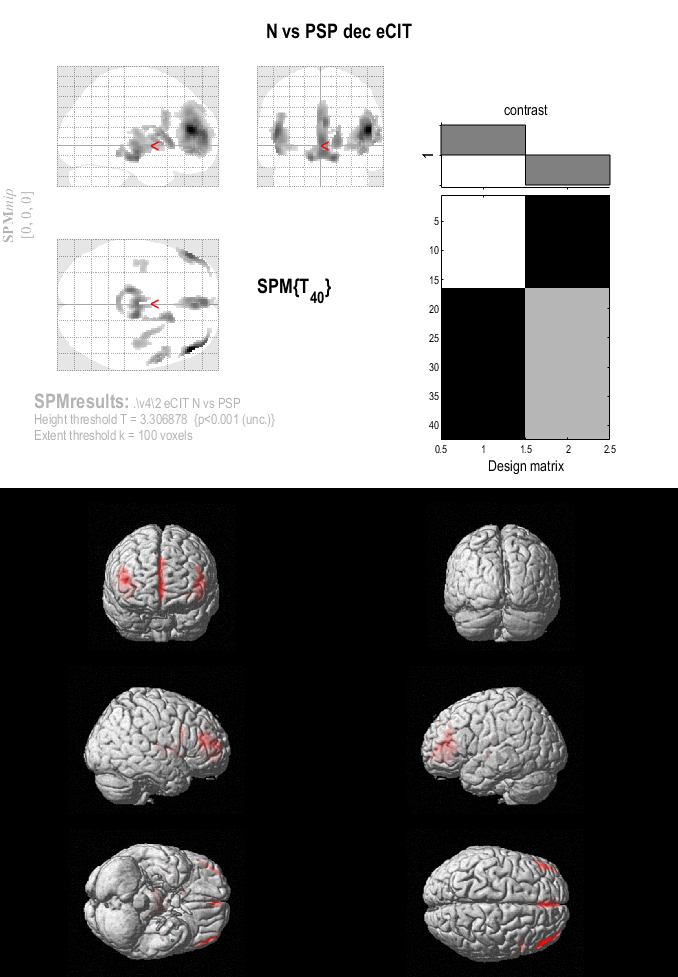
Fig 1. Perfusion PET: PSP vs healthy control
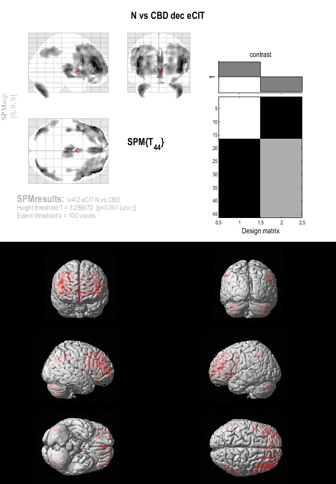
Fig 1. Perfusion PET: CBD vs healthy control
To cite this abstract in AMA style:
C. Lee, S. Kim, J. Ko, D. Doudet. Comparison of Regional Perfusion Patterns in Clinical Subtypes of PSP and CBD: Dual Phase [¹⁸F]FP CIT PET Study [abstract]. Mov Disord. 2026; 41 (suppl 1). https://www.mdsabstracts.org/abstract/comparison-of-regional-perfusion-patterns-in-clinical-subtypes-of-psp-and-cbd-dual-phase-%c2%b9%e2%81%b8ffp-cit-pet-study/. Accessed October 1, 2026.« Back to 2026 International Congress
MDS Abstracts - https://www.mdsabstracts.org/abstract/comparison-of-regional-perfusion-patterns-in-clinical-subtypes-of-psp-and-cbd-dual-phase-%c2%b9%e2%81%b8ffp-cit-pet-study/