
Category: Parkinson's disease: Neuroimaging
Objective: To investigate whether cerebellar peduncle microstructure is associated with autonomic dysfunction in Parkinson’s disease (PD).
Background: Autonomic dysfunction is a common and disabling non-motor manifestation of PD. Heart rate variability (HRV) provides a non-invasive marker of autonomic dysfunction. Emerging evidence suggests that the cerebellum participates in autonomic regulation, but the structural substrates linking cerebellar pathways to autonomic dysfunction in PD remain unclear.
Method: We analyzed data from the Prospective Assessments with Neuroimaging and Fluid Data Analysis in Parkinson’s Disease (PANDA) cohort, including 173 patients with PD (PwPD) and 31 healthy controls (HC). Autonomic symptoms were assessed using the SCOPA-AUT scale, and HRV parameters were derived from 24-hour electrocardiography. Diffusion MRI metrics of cerebellar peduncles were estimated using free-water diffusion model. Associations among HRV indices, SCOPA, and cerebellar peduncle integrity were examined using regression and mediation analyses adjusted for age and sex.
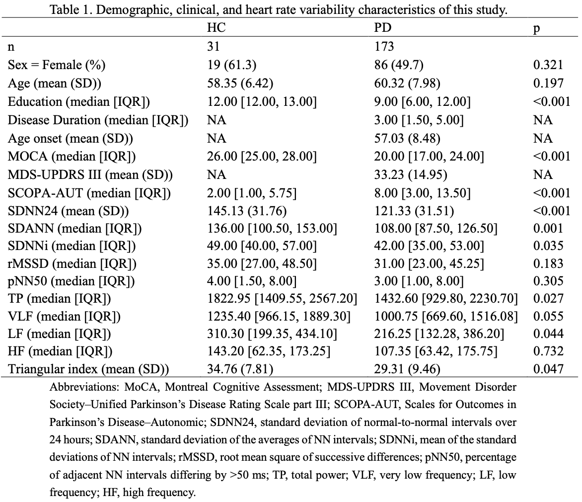
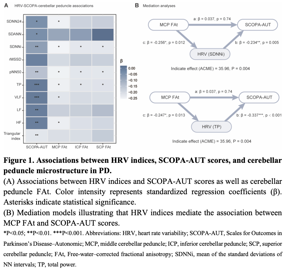
Results: PwPD showed greater autonomic symptom burden compared with HC (SCOPA median 8 vs 2, p<0.001) and reduced HRV measures (p<0.05). Lower HRV was associated with higher SCOPA scores, with total frequency-domain power (TP; p<0.001), VLF (p<0.001), LF (p=0.022), and HF (p=0.033) remaining significant after Bonferroni correction. Free-water–corrected FA (FAt) of the middle cerebellar peduncle (MCP) was significantly associated with multiple HRV indices, including SDNN24, SDNNi, pNN50, TP, VLF, and HF (all p<0.05). Similar associations were observed for inferior and superior cerebellar peduncles, with significant relationships between FAt and SDNNi, pNN50, and TP (all p<0.05). Mediation analysis demonstrated that SDNNi significantly mediated the association between MCP FAt and SCOPA (ACME=35.96, p=0.004), with similar mediating effects observed for TP, pNN50, VLF, and HF (all p<0.05).
Conclusion: Cerebellar peduncle microstructure is associated with autonomic dysfunction in PD. HRV may represent a physiological pathway linking cerebellar pathway integrity to autonomic symptom burden, supporting a potential role of cerebellar circuits in autonomic regulation
Table
Figure
To cite this abstract in AMA style:
RY. Yang, YUZ. Zhang. Cerebellar peduncle microstructure and autonomic dysfunction in Parkinson’s disease [abstract]. Mov Disord. 2026; 41 (suppl 1). https://www.mdsabstracts.org/abstract/cerebellar-peduncle-microstructure-and-autonomic-dysfunction-in-parkinsons-disease/. Accessed October 1, 2026.« Back to 2026 International Congress
MDS Abstracts - https://www.mdsabstracts.org/abstract/cerebellar-peduncle-microstructure-and-autonomic-dysfunction-in-parkinsons-disease/