
Objective: To investigate whole-cortex functional alterations in Parkinson’s disease patients with freezing of gait (PD-FOG) using transcranial magnetic stimulation combined with electroencephalography (TMS-EEG).
Background: Our Previous study linked PD-FOG to motor cortical disinhibition, but large-scale cortical dynamics remain unclear. TMS-EEG provides high temporal resolution and captures cortical responses by perturbing the primary motor cortex (M1) at rest. TMS-evoked potentials (TEPs) reflect cortical excitability, and event-related spectral perturbation (ERSP) captures oscillatory dynamics.
Method: Nineteen healthy controls (HCs), 18 PD patients without FOG (PD Non-FOG), and 34 PD patients with FOG (PD-FOG) were enrolled, including 16 with “off-period” FOG (OFF-FOG) and 18 with “levodopa-unresponsive” FOG (ONOFF-FOG). PD participants underwent TMS-EEG and gait assessments during medication OFF and ON states. Group differences in TEPs and ERSPs were analyzed in the OFF state, and correlations with gait parameters were examined. Dopaminergic effects were examined across FOG subtypes. 10-Hz repetitive TMS (rTMS) effects were evaluated in ONOFF-FOG.
Results: In OFF state, PD-FOG showed reduced N45 and enhanced P60 over motor cortex versus PD Non-FOG and HCs. In PD-FOG, N45 amplitude correlated positively with stride time coefficient of variation (CV), stride frequency CV, and double-support phase, and negatively with swing phase. P60 amplitude correlated positively with stride time CV. ERSP revealed increased beta-band synchronization in central, frontoparietal, and occipital regions in PD-FOG. Beta power in central/frontoparietal areas correlated positively with gait variability and negatively with stride length. Dopaminergic medication normalized N45, P60, and beta synchronization in OFF-FOG but not ONOFF-FOG. High-frequency rTMS improved gait performance in ONOFF-FOG patients and was accompanied by normalization of these neurophysiological abnormalities.
Conclusion: TEPs findings revealed local motor cortical disinhibition, while enhanced beta-band hypersynchrony in central, frontoparietal, and occipital regions reflected increased inhibition at a broader network level. These local and global abnormalities may interact to create a vulnerable motor network, contributing to FOG pathophysiology.
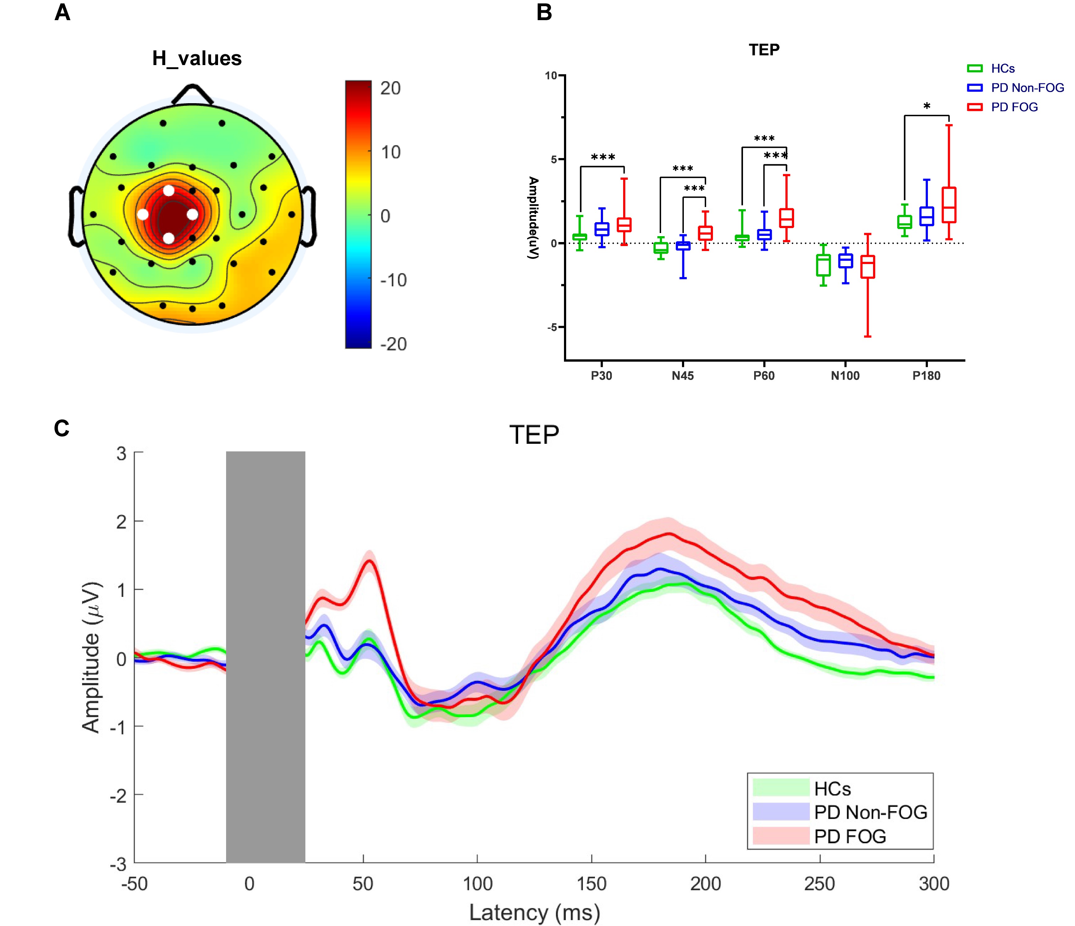
Figure1
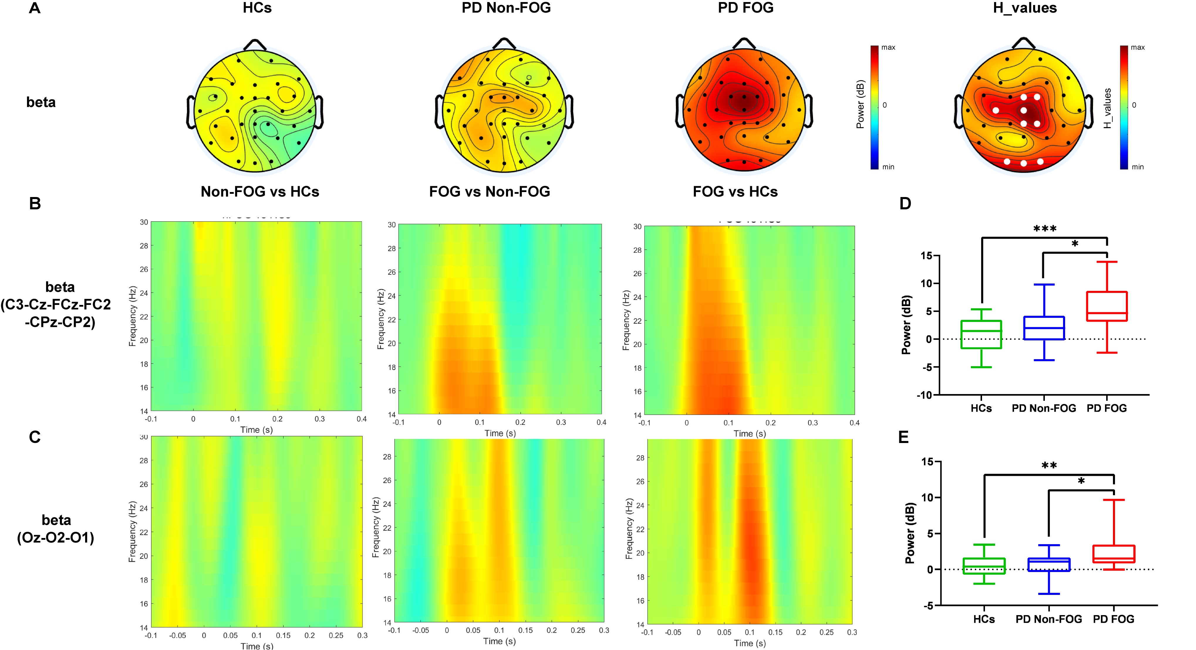
Figure2
To cite this abstract in AMA style:
HM. Sun, KZ. Zhang. Cortical Disinhibition and Beta Hypersynchrony underlying Freezing of Gait in Parkinson’s Disease: a TMS–EEG Study [abstract]. Mov Disord. 2026; 41 (suppl 1). https://www.mdsabstracts.org/abstract/cortical-disinhibition-and-beta-hypersynchrony-underlying-freezing-of-gait-in-parkinsons-disease-a-tms-eeg-study/. Accessed October 1, 2026.« Back to 2026 International Congress
MDS Abstracts - https://www.mdsabstracts.org/abstract/cortical-disinhibition-and-beta-hypersynchrony-underlying-freezing-of-gait-in-parkinsons-disease-a-tms-eeg-study/