
Category: Parkinson's disease: Neuroimaging
Objective: To investigate grey matter volume and white matter microstructural alterations in PRKN-PD using multimodal MRI and to compare these features with those observed in early-onset idiopathic PD (iEOPD) and healthy controls (HC).
Background: PRKN is the most common autosomal recessive causative gene for Parkinson’s disease (PD). Patients with PRKN-related PD (PRKN-PD) exhibit distinct pathological and clinical characteristics compared with idiopathic PD (iPD). However, differentiation between PRKN-PD and iPD based solely on clinical features remains challenging. Identifying imaging characteristics specific to PRKN-PD may therefore facilitate more accurate diagnosis, guide treatment strategies, and support outcome evaluation in clinical trials.
Method: Eighteen PD patients with bi-allelic PRKN mutations (PRKN-PD), 19 patients with early-onset PD without PD-causing mutations (iEOPD), and 23 age- and sex-matched healthy controls were included. All participants underwent clinical assessment and multimodal MRI. Grey matter structure was analyzed using voxel-based morphometry (VBM) and region-of-interest (ROI) volumetric analysis with FreeSurfer based on 3D-T1 images. Diffusion tensor imaging (DTI) was used to assess white matter microstructure. Fractional anisotropy (FA), radial diffusivity (RD), axial diffusivity (AD), and mean diffusivity (MD) were derived and analyzed using tract-based spatial statistics (TBSS).
Results: VBM analysis revealed significant differences in the bilateral caudate nucleus among the PRKN-PD, iEOPD, and HC groups (Fig.1). FreeSurfer analysis further demonstrated reduced bilateral caudate nucleus volume in the PRKN-PD group compared with both the HC (Left: p = 0.007; Right: p = 0.008) and iEOPD (Left: p = 0.019; Right: p = 0.039) groups, whereas no significant difference was observed between the iEOPD and HC groups. In white matter analyses, the PRKN-PD group showed reduced FA values in the corticospinal tract and superior longitudinal fasciculus compared with HC (Fig.2). In contrast, the iEOPD group exhibited significantly increased FA in multiple white matter tracts compared with HC (Fig.3), and no significant differences in FA, MD, AD, or RD were observed between the PRKN-PD and iEOPD groups.
Conclusion: Caudate nucleus atrophy may represent a relatively specific imaging feature of PRKN-PD. In addition, PRKN-PD may exhibit distinct white matter microstructural alterations.
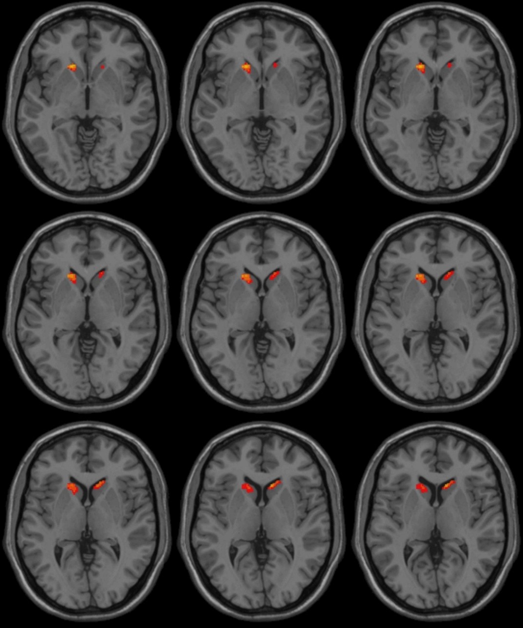
Fig.1
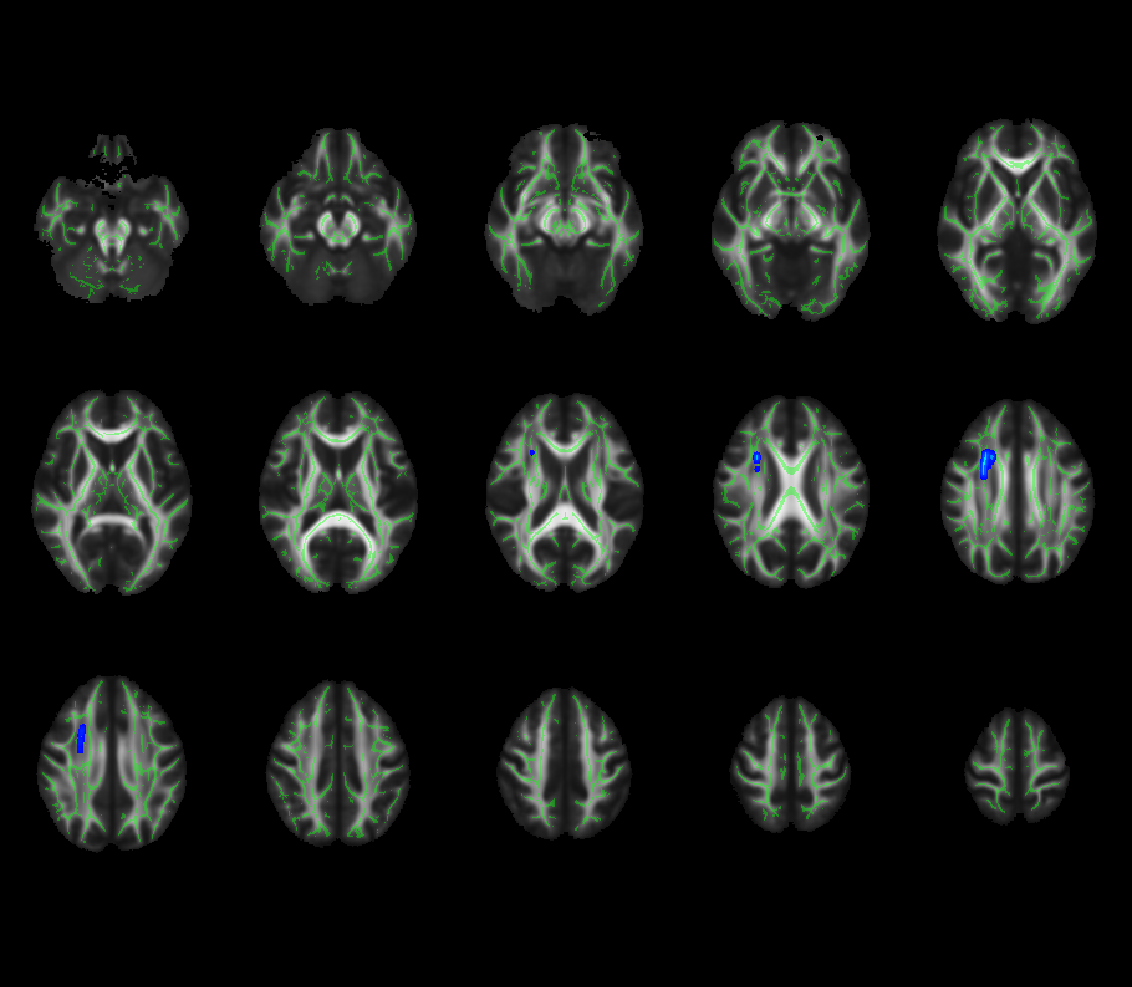
Fig.2
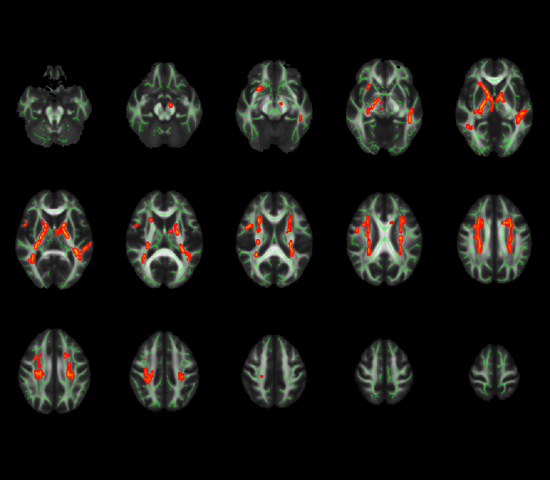
Fig.3
To cite this abstract in AMA style:
HT. Wang, F. Xie, B. Wang, W. Luo. Grey Matter Volume and White Matter Microstructural Alterations in PRKN-PD [abstract]. Mov Disord. 2026; 41 (suppl 1). https://www.mdsabstracts.org/abstract/grey-matter-volume-and-white-matter-microstructural-alterations-in-prkn-pd/. Accessed October 1, 2026.« Back to 2026 International Congress
MDS Abstracts - https://www.mdsabstracts.org/abstract/grey-matter-volume-and-white-matter-microstructural-alterations-in-prkn-pd/