
Category: MSA, PSP, CBS: Neuroimaging
Objective: To assess and quantify the functional brain networks in resting-state fMRI (rsfMRI) of patients with spinocerebellar ataxia type 12 (SCA 12) compared with healthy subjects.
Background: SCA12, caused by PPP2R2B CAG repeat expansion, explains with action tremor preceding ataxia, dysarthria, and cognitive decline (1). rsfMRI measures intrinsic brain connectivity via BOLD fluctuations, revealing network alterations in neurodegenerative diseases. SCA12 demonstrates cerebello-cortical functional disruptions despite milder structural changes (1).
Method: Genetically confirmed 52 SCA12 (male 34/female 18; age 43.66±9.00), along with 52 healthy controls (male 27/female 25; age 42.15±11.53) were enrolled. Clinical severity measured by the SARA score (11.14 ± 6.08).MRI data were acquired on a 3T Philips Ingenia scanner using a 32-channel head coil. 3D T1-weighted TFE sequence: TR=8.1 ms, TE=3.7 ms, FOV=250 mm, 1 mm isovoxel, 350 slices, flip angle=10°. rsfMRI EPI sequence: 220 dynamics, 35 axial slices, TR=2000 ms, TE=30 ms, FOV=230×230×140 mm, voxel=3×3×4 mm³ (8.7 min). Data analyzed using CONN software with default parameters (3).resting-state networks underwent standard preprocessing (realignment, co-registration to T1, normalization, denoising). Group difference identified (p<0.05 FDR voxel, p<0.05 FWE cluster).
Results: Seed-to-voxel analysis revealed significant network alterations in SCA12 versus healthy controls (Figure 1 and Table 1). The default mode network exhibited hypoconnectivity in cerebellar lobule IX (Figure 2). Sensorimotor networks showed hyperconnectivity in bilateral insular cortex, Heschl’s gyrus, and opercular regions in SCA12 patients (Figure 3). These distinctive alterations in sensory integration networks underscore SCA12’s unique tremor-dominant phenotype, differentiating it from classical ataxias characterized by predominant cerebellar pathology.
Conclusion: This rsfMRI study reveals distinct functional connectivity alterations in SCA12, with differential DMN hypoconnectivity and sensorimotor hyperconnectivity in the opercular/insular regions relative to controls. SCA12 demonstrates reorganized but relatively preserved networks, supporting its slower progression. These imaging biomarkers enable SCA12 to differentiate and predict functional prognosis.
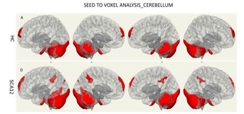
rsfMRI seed-based cerebellum connectivity
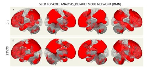
rsfMRI default mode network (DMN) connectivity
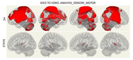
rsfMRI Sensory motor (SMN) connectivity
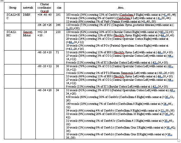
Resting State-Seed based analysis of SCA12
References: 1. Choubtum, L., Witoonpanich, P., Hanchaiphiboolkul, S., Bhidayasiri, R., Jitkritsadakul, O., Pongpakdee, S., Wetchaphanphesat, S., Boonkongchuen, P., & Pulkes, T. (2015). Analysis of SCA8, SCA10, SCA12, SCA17 and SCA19 in patients with unknown spinocerebellar ataxia: A Thai multicentre study. BMC Neurology, 15, 166. https://doi.org/10.1186/s12883-015-0425-y
2. Cocozza, S., Saccà, F., Cervo, A., Marsili, A., Russo, C. V., Giorgio, S. M. D. A., De Michele, G., Filla, A., Brunetti, A., & Quarantelli, M. (2015a). Modifications of resting state networks in spinocerebellar ataxia type 2. Movement Disorders : Official Journal of the Movement Disorder Society, 30(10), 1382–1390. https://doi.org/10.1002/mds.26284
3. Nieto-Castanon, A. (2022). Brain-wide connectome inferences using functional connectivity MultiVariate Pattern Analyses (fc- MVPA). PLoS Comput Biol 18(11): e1010634.
To cite this abstract in AMA style:
P. Pankaj, M. Kumar, S. Kumaran, A. Srivastava, A. Garg, R. Agrawal, A. Nehra, R. Rajan. Mapping Intrinsic Brain Network Connectivity Using Resting-State Functional MRI In Spinocerebellar Ataxia 12 In Comparison with Healthy Controls [abstract]. Mov Disord. 2026; 41 (suppl 1). https://www.mdsabstracts.org/abstract/mapping-intrinsic-brain-network-connectivity-using-resting-state-functional-mri-in-spinocerebellar-ataxia-12-in-comparison-with-healthy-controls/. Accessed October 1, 2026.« Back to 2026 International Congress
MDS Abstracts - https://www.mdsabstracts.org/abstract/mapping-intrinsic-brain-network-connectivity-using-resting-state-functional-mri-in-spinocerebellar-ataxia-12-in-comparison-with-healthy-controls/