
Category: Parkinson's disease: Neuroimaging
Objective: This study elucidates spatiotemporal functional connectivity of pure fatigue in early Parkinson’s disease (PD) and uncovering underlying gene expression profiles.
Background: Fatigue is a prevalent, debilitating non-motor symptom in early PD, yet its CNS mechanisms remain unclear. Isolate “pure” fatigue mechanisms in PD remains a critical gap. This study addresses this by dissecting the neural circuitry and molecular landscape of fatigue while controlling for confounders.
Method: Participants: 76 subjects: 31 early PD with pure fatigue (PD-F), 31 without fatigue (PD-NF), and 14 healthy controls (HC). Strict exclusion criteria removed depression, anxiety, sleep disorders, apathy, sarcopenia, and cognitive impairment.
Imaging: 3.0T rs-fMRI (medication-off/naïve). Calculated sFCD and dFCD.
Transcriptomics: Used JuSpace for neurotransmitter/cell-type correlation and Allen Human Brain Atlas for Gene Category Enrichment Analysis (GCEA).
Results: Clinical: PD-F showed greater motor/functional disability (MDS-UPDRS II/III) and higher FSS scores (5.58±0.95 vs 2.06±0.84, p<0.001) than PD-NF, with no significant differences in mood/cognition, confirming pure fatigue isolation.
Figure 1 illustrates reduced dFCD in left calcarine fissure and right precentral gyrus; shows negative correlation with FSS).
Molecular Correlations: Fatigue-related dFCD alterations spatially correlated with Dopamine D2 receptors (p=0.014), COX-1 (neuroinflammation, p=0.007), glucose metabolism (p=0.011), HDAC expression (epigenetics, p=0.027), and mitochondrial density (p=0.016).
Figure 2 visualizes spatial overlaps with D2, COX-1, glucose, HDAC, and mitochondria.
Gene Expression: GCEA revealed enrichment in protein modification/epigenetic regulation, RNA processing, glucose/lipid metabolism, neuroinflammation, blood-brain barrier function, and dopaminergic/cholinergic signaling.
Figure 3 Displays enriched GO categories: epigenetic regulation, RNA processing, energy metabolism, neuroinflammation, BBB function, receptor signaling.
Conclusion: Fatigue in early PD involves specific spatiotemporal connectivity abnormalities in visual/sensorimotor cortices, rooted in molecular pathologies like histone acetylation dysregulation, neuroinflammation (COX-1), and metabolic dysfunction.

Table 1: Clinical characteristics
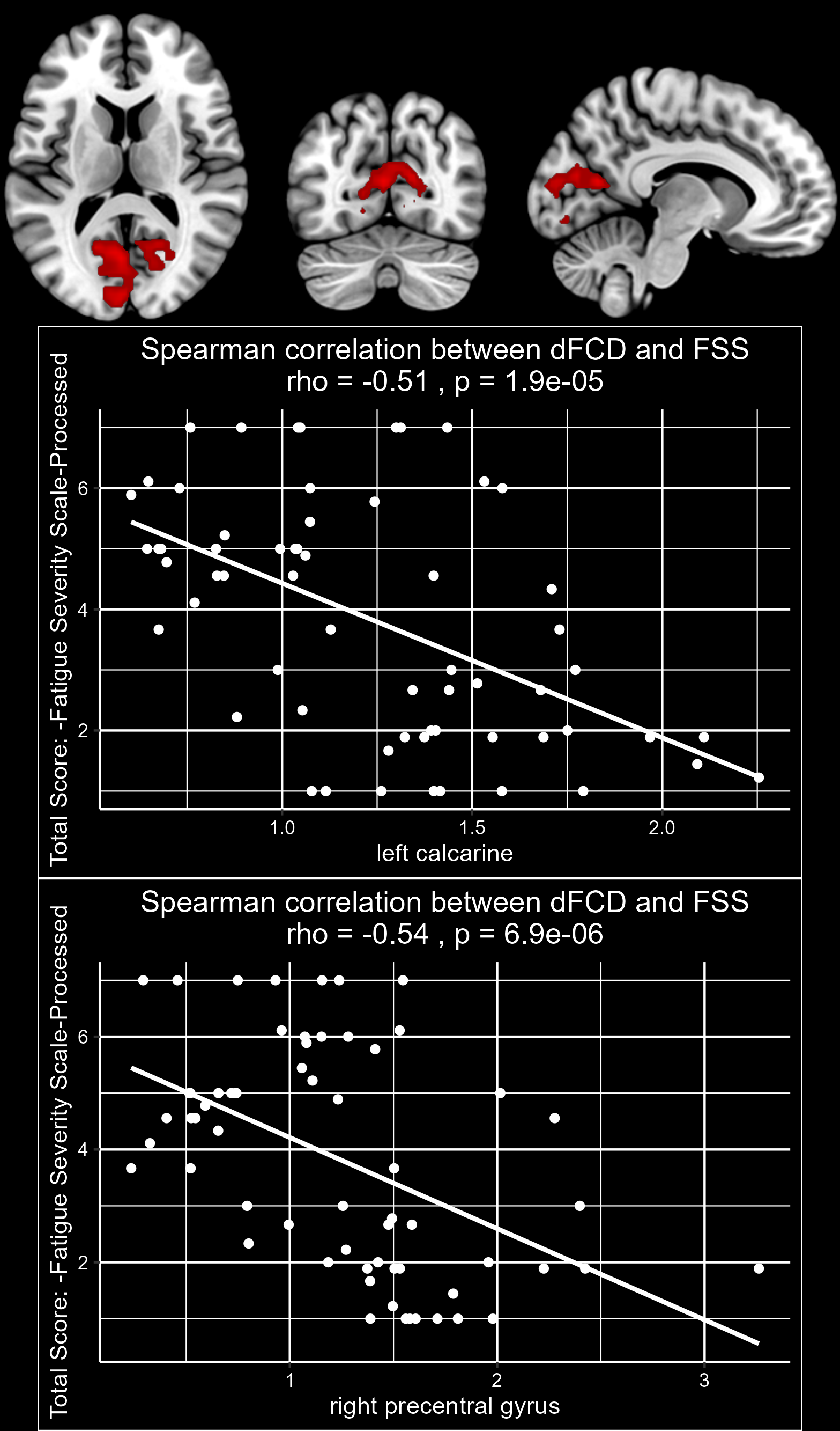
Figure 1: Fatigue-related dFCD in PD
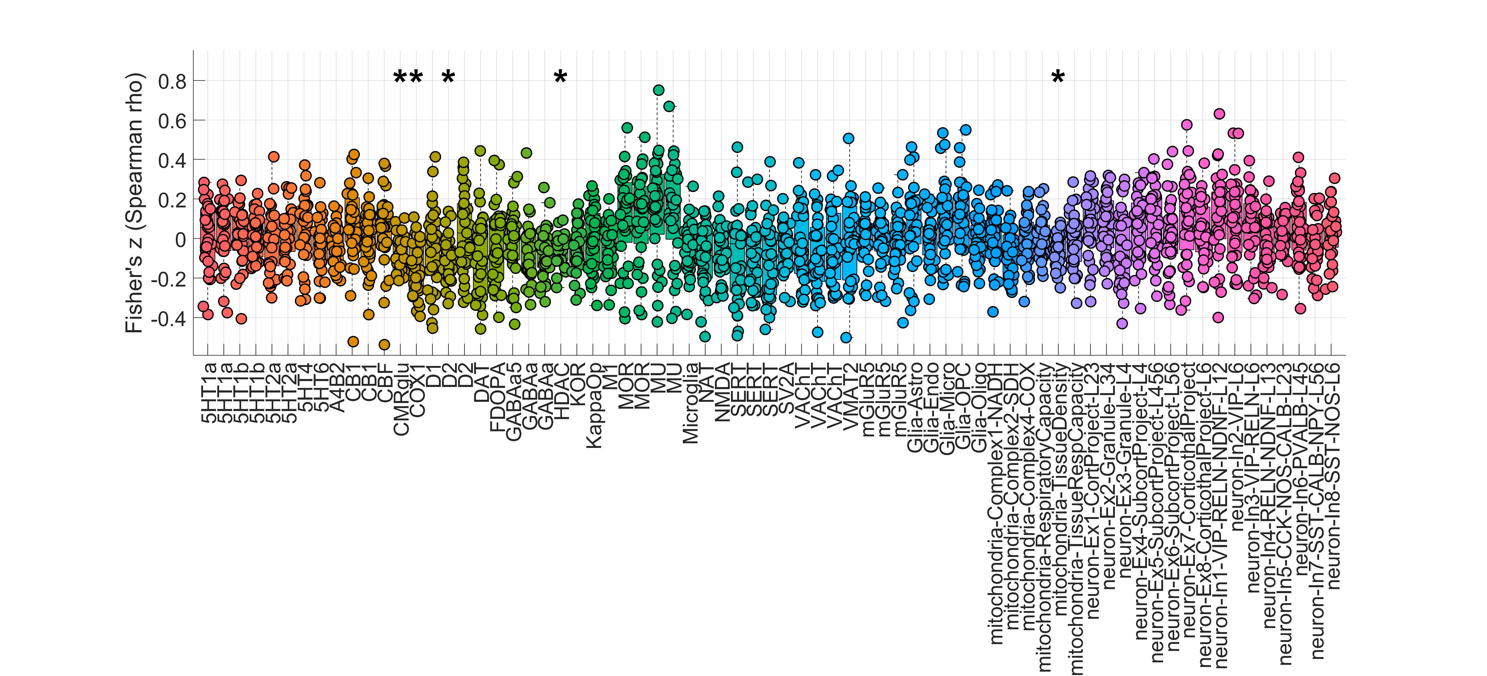
Figure 2
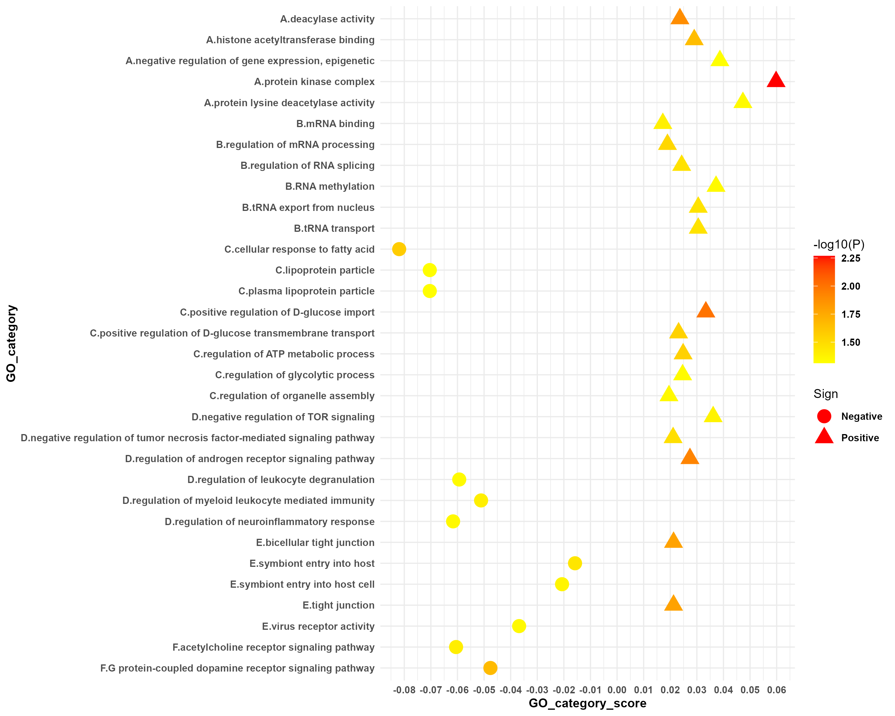
Figure 3: Gene categories
References: Reference
1. Chaudhuri A, Behan PO. Fatigue in neurological disorders. The Lancet. 2004;363(9413):978–988. doi: 10.1016/S0140-6736(04)15794-2.
2. Kluger BM, Herlofson K, Chou KL, et al. Parkinson’s disease‐related fatigue: A case definition and recommendations for clinical research. Mov Disord. 2016;31(5):625–631. doi: 10.1002/mds.26511.
3. Siciliano M, Trojano L, Santangelo G, De Micco R, Tedeschi G, Tessitore A. Fatigue in Parkinson’s disease: A systematic review and meta-analysis. Mov Disord Off J Mov Disord Soc. 2018;33(11):1712–1723. doi: 10.1002/mds.27461.
4. Ou R, Hou Y, Liu K, et al. Progression of Fatigue in Early Parkinson’s Disease: A 3-Year Prospective Cohort Study. Front Aging Neurosci. 2021;13:701906. doi: 10.3389/fnagi.2021.701906.
5. Elbers RG, Berendse HW, Kwakkel G. Treatment of Fatigue in Parkinson Disease. JAMA. 2016;315(21):2340. doi: 10.1001/jama.2016.5260.
6. Elbers RG, Verhoef J, Van Wegen EE, Berendse HW, Kwakkel G. Interventions for fatigue in Parkinson’s disease. Cochrane Movement Disorders Group, editor. Cochrane Database Syst Rev. 2015;2015(10). doi: 10.1002/14651858.CD010925.pub2.
7. Hou Y, Zhang L, Ou R, et al. Resting-state fMRI study on drug-naïve early-stage patients with parkinson’s disease and with fatigue. Parkinsonism Relat Disord. 2022;105:75–82. doi: 10.1016/j.parkreldis.2022.11.009.
8. Tessitore A, Giordano A, De Micco R, et al. Functional connectivity underpinnings of fatigue in “drug-naïve” patients with parkinson’s disease. Mov Disord Off J Mov Disord Soc. 2016;31(10):1497–1505. doi: 10.1002/mds.26650.
9. Shan A, Zhang H, Gao M, et al. Aberrant voxel‐based degree centrality and functional connectivity in parkinson’s disease patients with fatigue. CNS Neurosci Ther. 2023;29(9):2680–2689. doi: 10.1111/cns.14212.
10. Lu Z-H, Chen Y-K, Fu X-L, Liu Y-L, Qu J-F, Xiao W-M. Global cortical atrophy may be associated with fatigue in patients with parkinson’s disease. J Clin Neurosci. 2020;79:172–177. doi: 10.1016/j.jocn.2020.07.004.
11. Shen EH, Overly CC, Jones AR. The allen human brain atlas. Trends Neurosci. Elsevier BV; 2012;35(12):711–714. doi: 10.1016/j.tins.2012.09.005.
12. Lotter LD, Saberi A, Hansen JY, et al. Regional patterns of human cortex development correlate with underlying neurobiology. Nat Commun. 2024;15(1):7987. doi: 10.1038/s41467-024-52366-7.
13. Martins D, Giacomel A, Williams SCR, Turkheimer F, Dipasquale O, Veronese M. Imaging transcriptomics: Convergent cellular, transcriptomic, and molecular neuroimaging signatures in the healthy adult human brain. Cell Rep. 2021;37(13):110173. doi: 10.1016/j.celrep.2021.110173.
14. Postuma RB, Berg D, Stern M, et al. MDS clinical diagnostic criteria for parkinson’s disease: MDS-PD clinical diagnostic criteria. Mov Disord. 2015;30(12):1591–1601. doi: 10.1002/mds.26424.
15. Friedman JH, Alves G, Hagell P, et al. Fatigue rating scales critique and recommendations by the movement disorders society task force on rating scales for parkinson’s disease. Mov Disord. 2010;25(7):805–822. doi: 10.1002/mds.22989.
16. Goetz CG, Fahn S, Martinez‐Martin P, et al. Movement disorder society‐sponsored revision of the unified parkinson’s disease rating scale (MDS‐UPDRS): Process, format, and clinimetric testing plan. Mov Disord. 2007;22(1):41–47. doi: 10.1002/mds.21198.
17. Hoops S, Nazem S, Siderowf AD, et al. Validity of the MoCA and MMSE in the detection of MCI and dementia in parkinson disease. Neurology. 2009;73(21):1738–1745. doi: 10.1212/WNL.0b013e3181c34b47.
18. Schrag A, Barone P, Brown RG, et al. Depression rating scales in parkinson’s disease: Critique and recommendations. Mov Disord. 2007;22(8):1077–1092. doi: 10.1002/mds.21333.
19. Matza LS, Morlock R, Sexton C, Malley K, Feltner D. Identifying HAM‐a cutoffs for mild, moderate, and severe generalized anxiety disorder. Int J Methods Psychiatr Res. 2010;19(4):223–232. doi: 10.1002/mpr.323.
20. Stiasny‐Kolster K, Mayer G, Schäfer S, Möller JC, Heinzel‐Gutenbrunner M, Oertel WH. The REM sleep behavior disorder screening questionnaire—a new diagnostic instrument. Mov Disord. 2007;22(16):2386–2393. doi: 10.1002/mds.21740.
21. Muntean M-L, Benes H, Sixel-Döring F, et al. Clinically relevant cut-off values for the parkinson’s disease sleep scale-2 (PDSS-2): A validation study. Sleep Med. 2016;24:87–92. doi: 10.1016/j.sleep.2016.06.026.
22. Johns MW. A new method for measuring daytime sleepiness: The epworth sleepiness scale. Sleep. 1991;14(6):540–545. doi: 10.1093/sleep/14.6.540.
23. Chen L-K, Woo J, Assantachai P, et al. Asian working group for sarcopenia: 2019 consensus update on sarcopenia diagnosis and treatment. J Am Med Dir Assoc. 2020;21(3):300-307.e2. doi: 10.1016/j.jamda.2019.12.012.
24. Jost ST, Kaldenbach M, Antonini A, et al. Levodopa dose equivalency in parkinson’s disease: Updated systematic review and proposals. Mov Disord. 2023;38(7):1236–1252. doi: 10.1002/mds.29410.
25. Jia X-Z, Wang J, Sun H-Y, et al. RESTplus: An improved toolkit for resting-state functional magnetic resonance imaging data processing. Sci Bull. 2019;64(14):953–954. doi: 10.1016/j.scib.2019.05.008.
26. Liao W, Wu G-R, Xu Q, et al. DynamicBC : A MATLAB Toolbox for Dynamic Brain Connectome Analysis. Brain Connect. 2014;4(10):780–790. doi: 10.1089/brain.2014.0253.
27. Chen Y, Cui Q, Xie A, et al. Abnormal dynamic functional connectivity density in patients with generalized anxiety disorder. J Affect Disord. 2020;261:49–57. doi: 10.1016/j.jad.2019.09.084.
28. Huang M. Histone acetylation contributes to mitochondrial dysfunction and neuroinflammation in parkinson’s disease [Doctor of Philosophy]. [Ames (Iowa)]: Iowa State University; 2022. doi: 10.31274/td-20240329-388.
29. Dukart J, Holiga S, Rullmann M, et al. JuSpace : a tool for spatial correlation analyses of magnetic resonance imaging data with nuclear imaging derived neurotransmitter maps. Hum Brain Mapp. 2021;42(3):555–566. doi: 10.1002/hbm.25244.
30. Schaefer A, Kong R, Gordon EM, et al. Local-global parcellation of the human cerebral cortex from intrinsic functional connectivity MRI. Cereb Cortex. 2018;28(9):3095–3114. doi: 10.1093/cercor/bhx179.
31. Markello RD, Arnatkeviciute A, Poline J-B, Fulcher BD, Fornito A, Misic B. Standardizing workflows in imaging transcriptomics with the abagen toolbox. eLife. eLife Sciences Publications, Ltd; 2021;10. doi: 10.7554/elife.72129.
32. Hawrylycz MJ, Lein ES, Guillozet-Bongaarts AL, et al. An anatomically comprehensive atlas of the adult human brain transcriptome. Nature. Springer Science and Business Media LLC; 2012;489(7416):391–399. doi: 10.1038/nature11405.
33. Arloth J, Bader DM, Röh S, Altmann A. Re-annotator: Annotation pipeline for microarray probe sequences. Xue Y, editor. PLOS ONE. 2015;10(10):e0139516. doi: 10.1371/journal.pone.0139516.
34. Hawrylycz M, Miller JA, Menon V, et al. Canonical genetic signatures of the adult human brain. Nat Neurosci. 2015;18(12):1832–1844. doi: 10.1038/nn.4171.
35. Fulcher BD, Arnatkeviciute A, Fornito A. Overcoming false-positive gene-category enrichment in the analysis of spatially resolved transcriptomic brain atlas data. Nat Commun. Springer Science and Business Media LLC; 2021;12(1). doi: 10.1038/s41467-021-22862-1.
36. Arnatkevic̆iūtė A, Fulcher BD, Fornito A. A practical guide to linking brain-wide gene expression and neuroimaging data. NeuroImage. Elsevier BV; 2019;189:353–367. doi: 10.1016/j.neuroimage.2019.01.011.
37. Burt JB, Helmer M, Shinn M, Anticevic A, Murray JD. Generative modeling of brain maps with spatial autocorrelation. NeuroImage. 2020;220:117038. doi: 10.1016/j.neuroimage.2020.117038.
38. Cui S, Jiang P, Cheng Y, Cai H, Zhu J, Yu Y. Molecular mechanisms underlying resting-state brain functional correlates of behavioral inhibition. NeuroImage. 2023;283:120415. doi: 10.1016/j.neuroimage.2023.120415.
39. Zhou X, Xiang Y, Song T, et al. Characteristics of fatigue in parkinson’s disease: A longitudinal cohort study. Front Aging Neurosci. 2023;15:1133705. doi: 10.3389/fnagi.2023.1133705.
40. Tamraz JC, Outin-Tamraz C, Saban R. Mr imaging anatomy of the optic pathways. Radiol Clin North Am. 1999;37(1):1–36. doi: 10.1016/S0033-8389(05)70076-2.
41. Veréb D, Kovács MA, Antal S, et al. Modulation of cortical resting state functional connectivity during a visuospatial attention task in parkinson’s disease. Front Neurol. Frontiers Media SA; 2022;13. doi: 10.3389/fneur.2022.927481.
42. Moro E, Bellot E, Meoni S, et al. Visual dysfunction of the superior colliculus in de novo parkinsonian patients. Ann Neurol. Wiley; 2020;87(4):533–546. doi: 10.1002/ana.25696.
43. Murray MM, Thelen A, Thut G, Romei V, Martuzzi R, Matusz PJ. The multisensory function of the human primary visual cortex. Neuropsychologia. 2016;83:161–169. doi: 10.1016/j.neuropsychologia.2015.08.011.
44. Siciliano M, De Micco R, Trojano L, et al. The psychological correlates of fatigue in parkinson’s disease: contribution of maladaptive metacognitive beliefs. Parkinsonism Relat Disord. Elsevier BV; 2021;91:135–138. doi: 10.1016/j.parkreldis.2021.09.020.
45. Di Luzio P, Tarasi L, Silvanto J, Avenanti A, Romei V. Human perceptual and metacognitive decision-making rely on distinct brain networks. Pack C, editor. PLOS Biol. 2022;20(8):e3001750. doi: 10.1371/journal.pbio.3001750.
46. Benwell NM, Mastaglia FL, Thickbroom GW. Reduced functional activation after fatiguing exercise is not confined to primary motor areas. Exp Brain Res. 2006;175(4):575–583. doi: 10.1007/s00221-006-0573-9.
47. Cotter G, Salah Khlif M, Bird L, E Howard M, Brodtmann A, Egorova-Brumley N. Post-stroke fatigue is associated with resting state posterior hypoactivity and prefrontal hyperactivity. Int J Stroke. 2022;17(8):906–913. doi: 10.1177/17474930211048323.
48. Ghazanfari N, Van Waarde A, Dierckx RAJO, Doorduin J, De Vries EFJ. Is cyclooxygenase‐1 involved in neuroinflammation? J Neurosci Res. Wiley; 2021;99(11):2976–2998. doi: 10.1002/jnr.24934.
49. Teismann P, Tieu K, Choi D-K, et al. Cyclooxygenase-2 is instrumental in parkinson’s disease neurodegeneration. Proc Natl Acad Sci. Proceedings of the National Academy of Sciences; 2003;100(9):5473–5478. doi: 10.1073/pnas.0837397100.
50. Knott C, Stern G, Wilkin GP. Inflammatory regulators in parkinson’s disease: INOS, lipocortin-1, and cyclooxygenases-1 and -2. Mol Cell Neurosci. Elsevier BV; 2000;16(6):724–739. doi: 10.1006/mcne.2000.0914.
51. Teismann P, Ferger B. Inhibition of the cyclooxygenase isoenzymes COX-1 and COX-2 provide neuroprotection in the MPTP-mouse model of parkinson’s disease. Synapse. Wiley; 2001;39(2):167–174. doi: 10.1002/1098-2396(200102)39:2<167::aid-syn8>3.0.co;2-u.
52. Kumar A, Vashist A, Kumar P, Kalonia H, Mishra J. Potential role of licofelone, minocycline and their combination against chronic fatigue stress induced behavioral, biochemical and mitochondrial alterations in mice. Pharmacol Rep. Springer Science and Business Media LLC; 2012;64(5):1105–1115. doi: 10.1016/s1734-1140(12)70907-6.
53. Kim M-J, Lee J-H, Juarez Anaya F, et al. First-in-human evaluation of [11C]PS13, a novel PET radioligand, to quantify cyclooxygenase-1 in the brain. Eur J Nucl Med Mol Imaging. 2020;47(13):3143–3151. doi: 10.1007/s00259-020-04855-2.
54. Uzuegbunam BC, Rummel C, Librizzi D, Culmsee C, Hooshyar Yousefi B. Radiotracers for imaging of inflammatory biomarkers TSPO and COX-2 in the brain and in the periphery. Int J Mol Sci. 2023;24(24):17419. doi: 10.3390/ijms242417419.
55. Herlofson K, Heijnen CJ, Lange J, et al. Inflammation and fatigue in early, untreated parkinson’s disease. Acta Neurol Scand. 2018;138(5):394–399. doi: 10.1111/ane.12977.
56. Eidem LE, Birkeland E, Austdal M, et al. Fatigue in parkinson’s disease: A proteomic study of cerebrospinal fluid. Mov Disord. Wiley; 2024;39(4):749–751. doi: 10.1002/mds.29715.
57. Eidelberg D, Moeller JR, Dhawan V, et al. The metabolic topography of parkinsonism. J Cereb Blood Flow Metab. SAGE Publications; 1994;14(5):783–801. doi: 10.1038/jcbfm.1994.99.
58. Zhang L, Li T, Yuan Y, et al. Brain metabolic correlates of fatigue in parkinson’s disease: A PET study. Int J Neurosci. Informa UK Limited; 2018;128(4):330–336. doi: 10.1080/00207454.2017.1381093.
59. Sahbai S, Kauv P, Abrivard M, et al. Severe posterior hypometabolism but normal perfusion in a patient with chronic fatigue syndrome/myalgic encephalomyelitis revealed by PET/MRI. Eur J Nucl Med Mol Imaging. Springer Science and Business Media LLC; 2019;46(2):531–532. doi: 10.1007/s00259-018-4229-3.
60. Tanaka M, Watanabe Y. Reduced energy utilization in the brain is a feature of an animal model of fatigue. Int J Neurosci. Informa UK Limited; 2008;118(5):683–692. doi: 10.1080/00207450701242974.
61. Zhang Y, Zhang Z, Yu Q, et al. Replicating human characteristics: A promising animal model of central fatigue. Brain Res Bull. Elsevier BV; 2024;212:110951. doi: 10.1016/j.brainresbull.2024.110951.
62. Wey H-Y, Gilbert TM, Zürcher NR, et al. Insights into neuroepigenetics through human histone deacetylase PET imaging. Sci Transl Med. 2016;8(351). doi: 10.1126/scitranslmed.aaf7551.
63. Catanese MC, Klingl YE, Gilbert TM, et al. Chemoproteomics sheds light on epigenetic targets of [11 C]martinostat in the human brain. ACS Chem Neurosci. 2025;16(4):723–731. doi: 10.1021/acschemneuro.4c00781.
64. Park G, Tan J, Garcia G, Kang Y, Salvesen G, Zhang Z. Regulation of histone acetylation by autophagy in parkinson disease. J Biol Chem. 2016;291(7):3531–3540. doi: 10.1074/jbc.M115.675488.
65. Gebremedhin KG, Rademacher DJ. Histone h3 acetylation in the postmortem parkinson’s disease primary motor cortex. Neurosci Lett. 2016;627:121–125. doi: 10.1016/j.neulet.2016.05.060.
66. Shvedunova M, Akhtar A. Modulation of cellular processes by histone and non-histone protein acetylation. Nat Rev Mol Cell Biol. 2022;23(5):329–349. doi: 10.1038/s41580-021-00441-y.
67. Charidemou E, Kirmizis A. A two-way relationship between histone acetylation and metabolism. Trends Biochem Sci. 2024;49(12):1046–1062. doi: 10.1016/j.tibs.2024.10.005.
68. Di Vico IA, Moretto M, Tamanti A, et al. Molecular‐informed network analysis unveils fatigue‐related functional connectivity in parkinson’s disease. Mov Disord. Wiley; 2025; doi: 10.1002/mds.30214.
69. Kaasinen V, Vahlberg T, Stoessl AJ, Strafella AP, Antonini A. Dopamine receptors in parkinson’s disease: A meta‐analysis of imaging studies. Mov Disord. 2021;36(8):1781–1791. doi: 10.1002/mds.28632.
To cite this abstract in AMA style:
Y. Xiao, S. Wang, Y. Hou, R. Ou, C. Li, W. Song, X. Shi, X. Zhu, H. Shang. The Transcriptomic, Neurotransmitter, and Cell Type Signatures Underlying Pure Fatigue in Early-stage Parkinson’s Disease [abstract]. Mov Disord. 2026; 41 (suppl 1). https://www.mdsabstracts.org/abstract/the-transcriptomic-neurotransmitter-and-cell-type-signatures-underlying-pure-fatigue-in-early-stage-parkinsons-disease/. Accessed October 1, 2026.« Back to 2026 International Congress
MDS Abstracts - https://www.mdsabstracts.org/abstract/the-transcriptomic-neurotransmitter-and-cell-type-signatures-underlying-pure-fatigue-in-early-stage-parkinsons-disease/