
Category: Autoimmune Movement Disorders
Objective: To identify neuronal antibodies in patients with unexplained chorea.
Background: In recent years, autoimmune causes of chorea are increasingly recognized and the number of neuronal antibodies associated with this clinical phenotype is growing. Importantly, not all these patients exhibit clear signs of an autoimmune etiology: disease course can be chronic rather than subacute and ancillary investigations (MRI, lumbar puncture) do not always show signs of inflammation.
Method: We retrospectively identified patients with unexplained chorea not suspected of an autoimmune cause seen at the outpatient clinics of three movement disorder tertiary referral centers (cohort A; n=56). All patients had previously undergone brain MRI to rule out structural causes, routine blood tests and genetic testing for Huntington’s disease, but no extensive autoimmune diagnostics. Serum and – if available – cerebrospinal fluid (CSF) of these patients was tested for neuronal antibodies by immunohistochemistry (IHC), immunoblot, cell-based assays (CBA) and hippocampal live neurons, based on clinical information. Secondly, we reviewed all patients with chorea (with or without additional features) in whom an autoimmune cause was part of the differential diagnosis and of whom blood or CSF was sent for antibody testing to our laboratory, the national referral center for neuronal antibody diagnostics (cohort B; n=86). These patient samples were tested with the same techniques.
Results: We found antibodies in 3/56 (5%; one of each of anti-LGI1, anti-CV2/CRMP5 and anti-KLHL11) of cohort A [figure 1]. All these patients had a slowly progressive disease course over years [table1]. In cohort B, we identified 12/86 antibody-positive cases (14%): five with anti-IgLON5 disease, two with other antibodies (one anti-GlyR, one anti-GABABR) and five probable cases with positive IHC but yet unidentified antibody target (additional investigations pending) [figure 1].
Conclusion: Neuronal antibody testing has a relevant diagnostic yield (5-14%) in patients with unexplained chorea and should be considered in all chorea cases without a clear structural or genetic cause. Slow disease progression and absence of CSF pleocytosis do not rule out autoimmune causes of chorea and should not preclude a patient from antibody testing, especially because of its therapeutic implications.
Abstract previously presented (poster) during the London Encephalitis Conference on December 4th 2025.
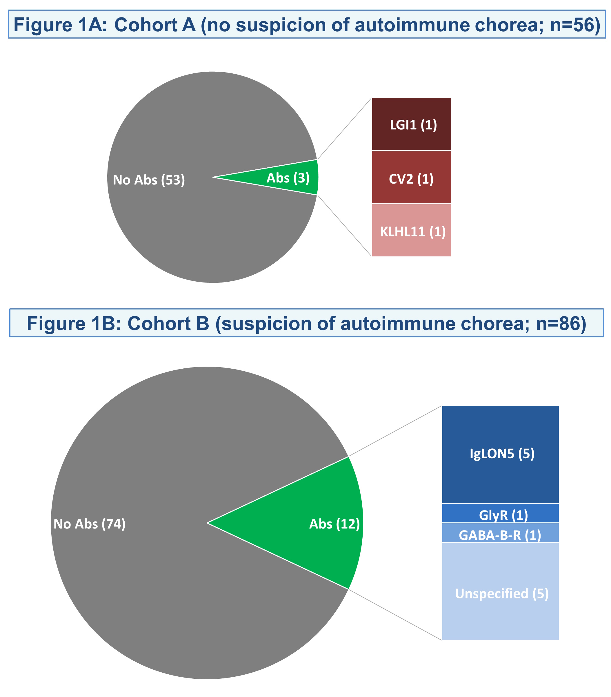
Antibody-Positive Patients in Each Cohort
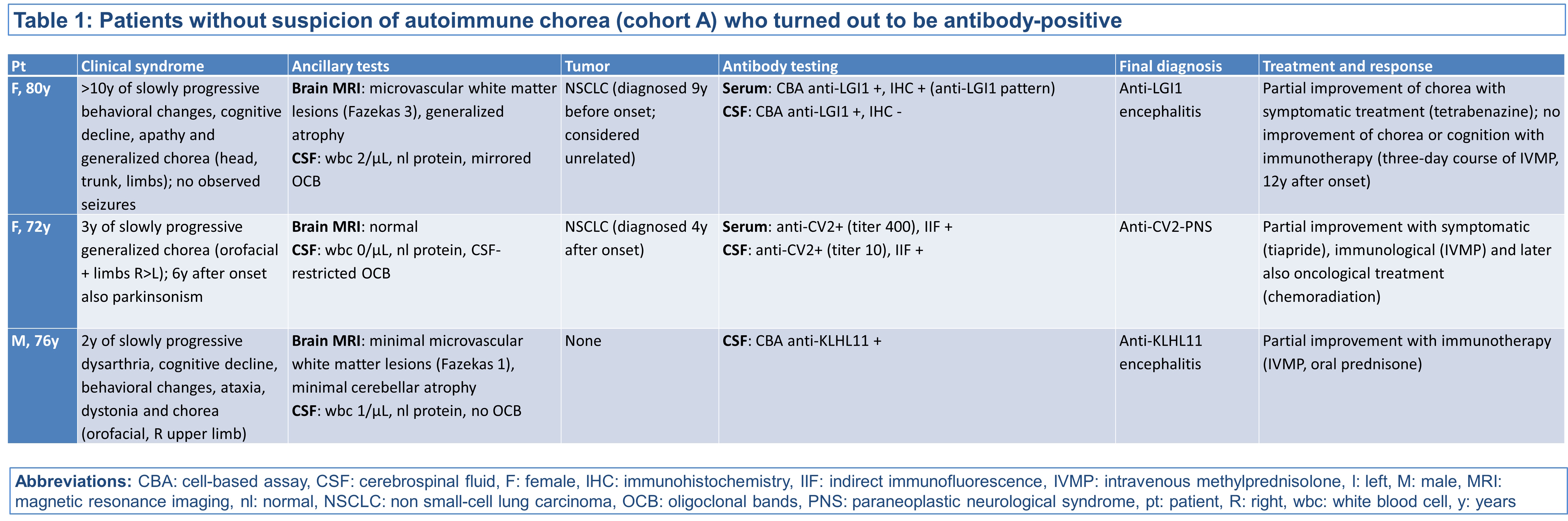
Details on Antibody-Positive Patients in Cohort A
To cite this abstract in AMA style:
J. Kerstens, J. de Vries, M. van Coevorden-Hameete, S. Franken, M. Nagtzaam, S. Veenbergen, E. Verhagen, P. Sillevis Smitt, S. de Bot, M. Oosterloo, A. Boon, M. Titulaer. “Unexplained” Chorea Explained by Neuronal Antibodies: A Dutch Cohort Study [abstract]. Mov Disord. 2026; 41 (suppl 1). https://www.mdsabstracts.org/abstract/unexplained-chorea-explained-by-neuronal-antibodies-a-dutch-cohort-study/. Accessed October 1, 2026.« Back to 2026 International Congress
MDS Abstracts - https://www.mdsabstracts.org/abstract/unexplained-chorea-explained-by-neuronal-antibodies-a-dutch-cohort-study/