
Objective: To develop and evaluate an integrated, deep learning (DL)-enhanced multimodal MRI classification model combining nigrosome-1 (N1), neuromelanin (NM), free-water DTI (FW-DTI) and morphometry (MORPHO) for Parkinsonian disorder subtyping at clinical presentation.
Background: Differentiating idiopathic Parkinson’s disease (iPD) from atypical (aPD) and secondary parkinsonism (sPD) at presentation is clinically challenging. Advanced MRI markers of nigral and brain structural integrity changes such as N1, NM, FW-DTI and MORPHO show individual diagnostic promise1-3, but their combined utility in real-world classification remains unknown.
Method: Brain MRI from 364 patients with final clinical diagnoses of iPD (n=219), aPD (n=37), and sPD (n=108) at 3-year follow-up were analysed (Table 1). Standardised 3T MRI (Siemens) included T1 MPRAGE, DTI, and high-resolution midbrain N1 and NM sequences. Brain morphometry was derived using FastSurfer4. N1 was assessed using two proprietary DL models on susceptibility map weighted imaging2. NM hyperintensity in the substantia nigra (SN) was segmented using MERIT, a vision transformer model trained on expert manual labels5, yielding intensity, contrast, volume, and shape metrics. FW maps were derived via the MarkVCID pipeline6, with FW values extracted from SN, subcortical nuclei and cerebellar lobules. All features were standardised and adjusted for age and sex; dimensionality reduction was applied to NM, FW-DTI and MORPHO features. Logistic regression with 5-fold cross-validation was used on an 80/20 stratified train-test split, evaluating 4 feature sets: (1) N1-NM, (2) FW-DTI, (3) MORPHO and (4) combined, across three classification tasks: iPD vs sPD, iPD vs aPD, and iPD vs non-PD (aPD+sPD).
Results: For iPD vs sPD, N1-NM achieved the best accuracy (89.3%); adding FW-DTI or MORPHO did not improve performance. For iPD vs aPD, N1-NM alone reached 71.1%, FW-DTI 75.6%, MORPHO 75.0%, and combining all three improved accuracy to 89.7%. For iPD vs non-PD, N1-NM reached 69.4%, FW-DTI 63.5%, and MORPHO 67.2%; combining N1-NM with MORPHO yielded the best performance at 80.4%.
Conclusion: N1-NM, FW-DTI, and MORPHO are complementary: N1-NM excels in iPD vs sPD discrimination, FW-DTI and MORPHO aid iPD vs aPD differentiation, and combining N1-NM with MORPHO optimises complex real-world iPD vs non-PD classification.
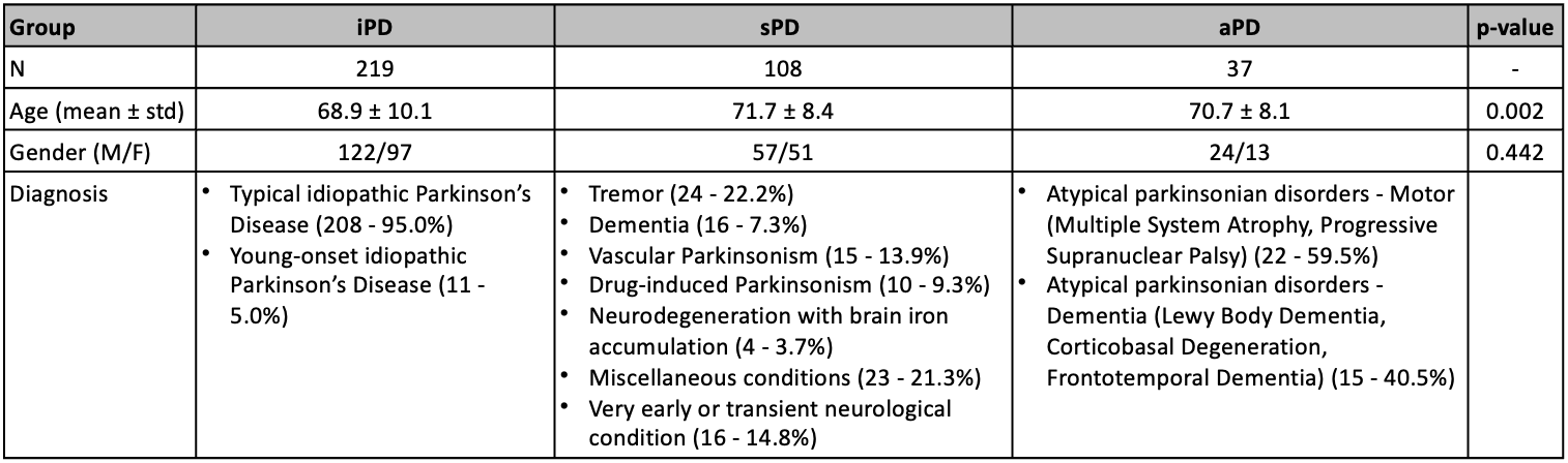
Table 1. Study demographics.
References: 1. Chau MT, Todd G, Wilcox R, et al. Diagnostic accuracy of the appearance of nigrosome-1 on magnetic resonance imaging in Parkinson’s disease: a systematic review and meta-analysis. Parkinsonism Relat Disord 2020;78:12–20. doi:10.1016/j.parkreldis.2020.07.002.
2. Welton T, Hartono S, Lee WL, et al. Classification of Parkinson’s disease by deep learning on midbrain MRI. Front Aging Neurosci 2024;16:1425095. doi:10.3389/fnagi.2024.1425095.
3. Vaillancourt DE, Barmpoutis A, Wu SS, et al. Automated imaging differentiation for parkinsonism. JAMA Neurol 2025;82:495–505. doi:10.1001/jamaneurol.2025.0112.
4. Henschel L, Conjeti S, Estrada S, et al. FastSurfer – A fast and accurate deep learning based neuroimaging pipeline. Neuroimage 2020 Oct 1:219:117012. doi: 10.1016/j.neuroimage.2020.117012.
5. Hartono S, Chen RC, Welton T, et al. Quantitative iron-neuromelanin MRI associates with motor severity in Parkinson’s disease and matches radiological disease classification. Front Aging Neurosci 2023;15:1287917. doi:10.3389/fnagi.2023.1287917.
6. Lu H, Kashani AH, Arfanakis K, et al. MarkVCID cerebral small vessel consortium: II. Neuroimaging protocols. Alzheimers Dement 2021;17:716–725. doi:10.1002/alz.12216
To cite this abstract in AMA style:
S. Hartono, H. Ulfah, Q. Sun, C. Liu, D. Patidar, P. Seow, P. Chai, Q. Lyu, R. Chen, E. Tan, L. Chan. Deep Learning-Enhanced Multimodal MRI for Real-World Differentiation of Parkinsonian Disorders [abstract]. Mov Disord. 2026; 41 (suppl 1). https://www.mdsabstracts.org/abstract/deep-learning-enhanced-multimodal-mri-for-real-world-differentiation-of-parkinsonian-disorders/. Accessed October 1, 2026.« Back to 2026 International Congress
MDS Abstracts - https://www.mdsabstracts.org/abstract/deep-learning-enhanced-multimodal-mri-for-real-world-differentiation-of-parkinsonian-disorders/