
Objective: To illustrate the clinical utility of postoperative DBS lead reconstruction and image-guided programming in genetic dystonia
Background: DBS of the globus pallidus internus (GPi) is an established therapy for generalized dystonia, but inaccurate electrode placement or suboptimal programming may limit outcomes. Imaging-based reconstruction allows precise electrode localization and may optimize programming.
Method: We describe three patients with genetically confirmed DYT-TOR1A dystonia who underwent DBS surgery at a tertiary public Brazilian center. Postoperative lead reconstructions using Lead-DBS (MATLAB) and StimView (Brainlab) were performed to evaluate electrode positioning and guide programming adjustments.
Results: Case 1: A 12-year-old boy with a 4-year history of progressive generalized severe dystonia (BFMDRS-M 88.5) underwent GPi-DBS three years after symptom onset without clinical improvement. Lead reconstruction demonstrated correct positioning of the left electrode but misplacement of the right side (figure 1A). Revision surgery was performed. Three months after reimplantation, the BFMDRS-M improved to 31 (64.7%). Appropriate new lead positioning was confirmed by imaging (figure 1B). Case 2: An 11-year-old girl with generalized dystonia (BFMDRS-M 76.5) underwent GPi-DBS and improved to BFMDRS-M 14 (81.6% at 3 months). Lead reconstruction guided stimulation toward GPi somatotopic regions corresponding to the lower limbs, enabling rapid parameter optimization (figure 2A and 2B). Case 3: A 27-year-old man with DYT-TOR1A dystonia underwent GPi-DBS surgery and developed intraoperative thalamic hemorrhage during the procedure, requiring interruption of surgery. One year later, bilateral STN-DBS was performed, improving BFMDRS-M from 76 to 23 (69.7%). High-amplitude stimulation on left. lead caused dysarthria. Lead reconstruction revealed a lateral left electrode position (figure 3). Bipolar configuration with lower pulse widths values reduced dysarthria while maintaining motor improvement.
Conclusion: Postoperative DBS lead reconstruction provides anatomical information to guide programming and clinical decision-making. Imaging-guided analysis enables detection of electrode misplacement, targeting of GPi somatotopic regions, and reduction of stimulation-related adverse effects. Systematic reconstruction may improve programming strategies and clinical outcomes in dystonia
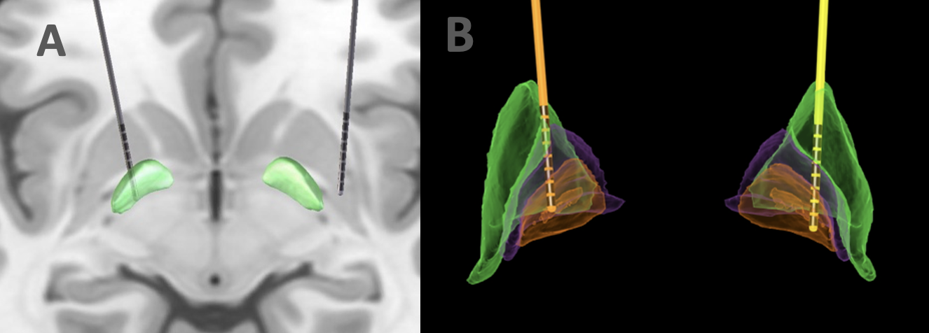
A. Lead misplacement. B. Correct GPi placement.
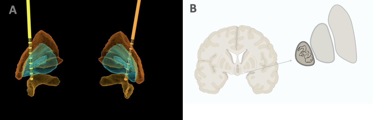
A. GPi lead reconstruction. B. GPi somatotopy.
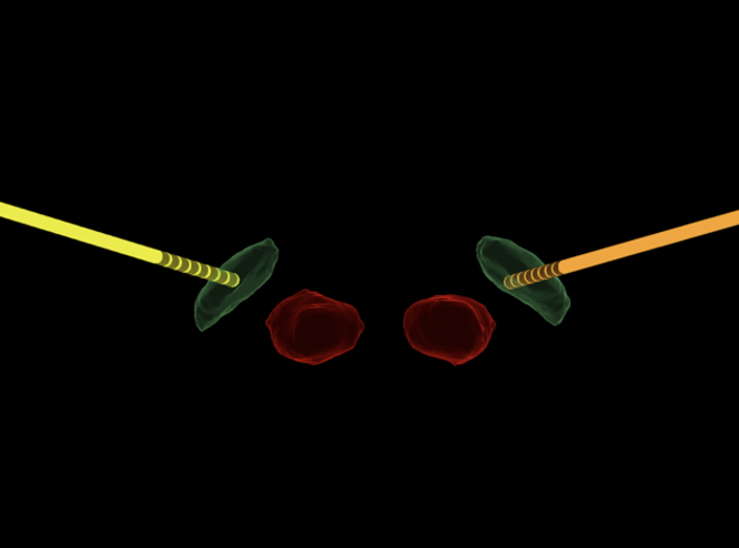
STN-DBS lead reconstruction with lateral left lead
References: Fan H, Zheng Z, Yin Z, Zhang J and Lu G (2021) Deep Brain Stimulation Treating Dystonia: A Systematic Review of Targets, Body Distributions and Etiology Classifications. Front. Hum. Neurosci. 15:757579. doi: 10.3389/fnhum.2021.757579
To cite this abstract in AMA style:
M. Soares, J. Nogueira, J. Gomes, M. Bezerra. Image-Guided DBS Programming in Dystonia: Experience from a Tertiary Public Brazilian Center [abstract]. Mov Disord. 2026; 41 (suppl 1). https://www.mdsabstracts.org/abstract/image-guided-dbs-programming-in-dystonia-experience-from-a-tertiary-public-brazilian-center/. Accessed October 1, 2026.« Back to 2026 International Congress
MDS Abstracts - https://www.mdsabstracts.org/abstract/image-guided-dbs-programming-in-dystonia-experience-from-a-tertiary-public-brazilian-center/