
Objective: To investigate whether the association between orthostatic blood pressure changes and structural neuroimaging markers of chronic cerebral hypoperfusion and small vessel disease differ by sex in patients with Parkinson’s disease (PD).
Background: Orthostatic hypotension (OH) is one of the most prevalent and debilitating non-motor symptoms in PD, often leading to episodic cerebral hypoperfusion. Although previous studies have suggested potential sex differences in the clinical manifestation of OH, whether the structural damage induced by OH-related blood pressure fluctuations also exhibit sex-specific patterns remains largely unexplored.
Method: This cross-sectional study included 217 patients with PD (103 women) from a prospective cohort. Orthostatic BP drops (ΔSBP, ΔDBP) and the presence of OH were assessed. Multi-modal MRI was utilized to quantify neuroimaging markers associated with cerebral hypoperfusion, including white matter hyperintensity (WMH), perivascular spaces (PVS), free-water (FW), peak width of skeletonized mean diffusivity (PSMD), and diffusivity along the perivascular spaces (DTI-ALPS) index. Sex-stratified linear regression models were applied.
Results: Baseline characteristics revealed a significantly higher prevalence of OH in men compared to women [table1], accompanied by more pronounced orthostatic drops in both SBP and DBP [figure1]. Linear model indicated distinct associations between orthostatic BP metrics and neuroimaging markers [table2] . In men, the presence of OH was significantly associated with increased total WMH volume. Furthermore, higher orthostatic BP drops in men associated with reduced basal ganglia PVS volumes and a lower DTI-ALPS index, suggesting potential impaired glymphatic and perivascular dynamics. In contrast, women did not show similar trend in most MRI markers. Notably, formal interaction models did not reach statistical significance for most sex-by-ΔSBP interactions.
Conclusion: Orthostatic blood pressure decline is associated with neuroimaging markers of small vessel disease and glymphatic impairment predominantly in men with PD. These findings suggest a potential sex-specific vulnerability to autonomic-mediated cerebral hypoperfusion. Our results highlight the importance of sex-stratified monitoring of orthostatic BP to mitigate potential cerebrovascular burden and neurodegenerative progression in PD patients.
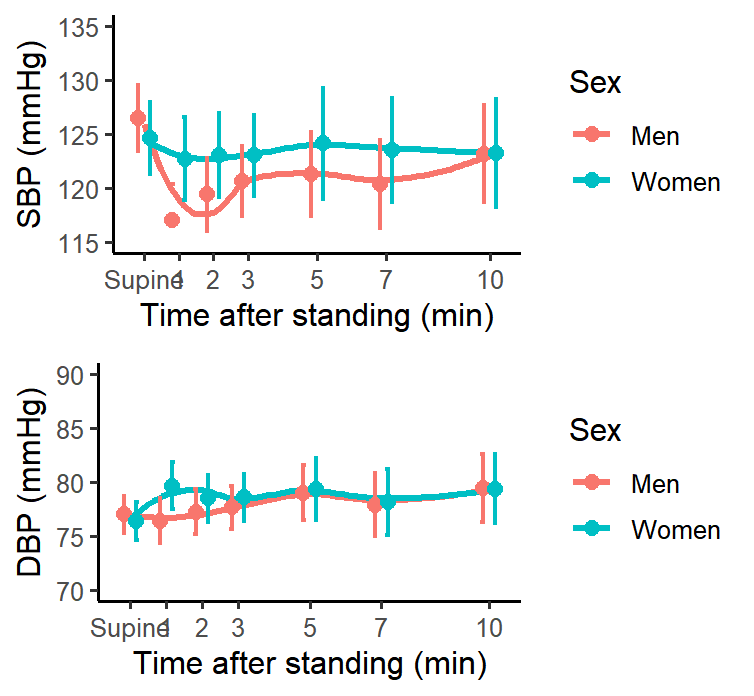
Figure 1. Orthostatic assessment
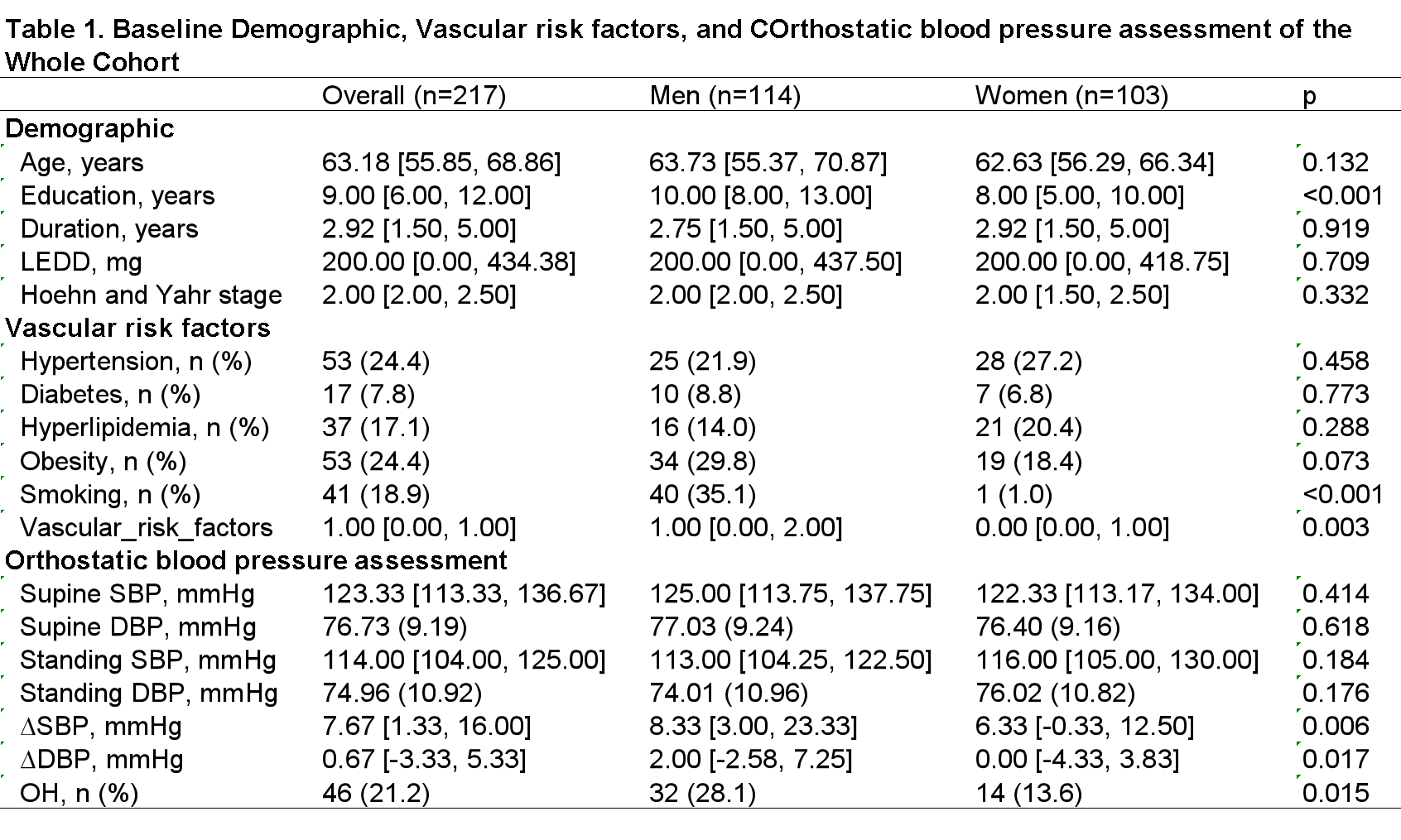
Table 1. Basic Characteristics
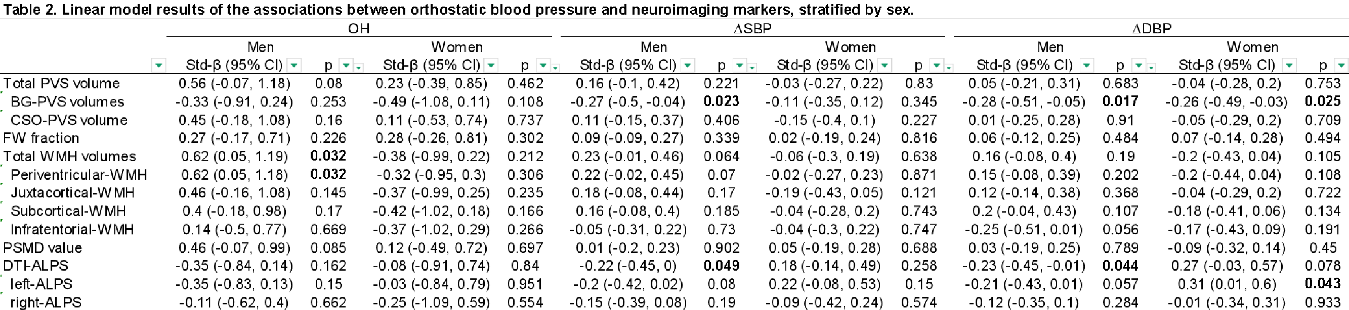
Table 2. Linear model results
To cite this abstract in AMA style:
Z. Li, X. Cui, S. Rong, R. Yang, M. Cai, Y. Zhang. Sex-Specific Associations Between Orthostatic Hypotension and Cerebral Hypoperfusion Injury in Parkinson’s Disease [abstract]. Mov Disord. 2026; 41 (suppl 1). https://www.mdsabstracts.org/abstract/sex-specific-associations-between-orthostatic-hypotension-and-cerebral-hypoperfusion-injury-in-parkinsons-disease/. Accessed October 1, 2026.« Back to 2026 International Congress
MDS Abstracts - https://www.mdsabstracts.org/abstract/sex-specific-associations-between-orthostatic-hypotension-and-cerebral-hypoperfusion-injury-in-parkinsons-disease/