
Category: Parkinson's disease: Neuroimaging
Objective: To develop and validate a T1-weighted structural MRI–based machine learning model for differentiating Parkinsonism at early-to-mid stage and generalize the model to routine anisotropic 2-dimensional (2D) clinical MRI.
Background: Multimodal research-grade magnetic resonance imaging (MRI) shows high diagnostic potential for differentiating Parkinson disease (PD) from multiple system atrophy (MSA). However, its clinical translation is limited by cost, acquisition time, and processing burden. A generalizable model for routine clinical MRI is needed.
Method: This multicenter cohort study was conducted from August 2018 to November 2025 across 5 clinical centers (China), with additional data from the Parkinson’s Progression Markers Initiative. Participants included healthy controls (HC), patients with PD, MSA, and idiopathic rapid eye movement sleep behavior disorder (iRBD) – a prodromal stage of PD. Patients were clinically diagnosed according to the established criteria and limited to early-to-mid disease stage. Participants were organized into a hierarchical discovery–verification–translation framework: Discovery, External Research-Grade, Exploratory iRBD, and External Clinical-MRI Cohort. Model parameters were fixed prior to external validation.
Results: Of 1053 screened individuals, 969 were included (mean [SD] age, 63.1 [8.7] years; 527 male [54.4%]; PD, 423; MSA, 367; iRBD, 107; HC, 72). In the internal testing set, the multimodal benchmark model achieved an Area under the receiver operating characteristic curve (AUROC) of 0.98 (95% CI, 0.94–1.00) for multiclass discrimination (HC vs PD vs MSA). A streamlined unimodal T1-Top20 model achieved an AUROC of 0.95 (95% CI, 0.89–1.00). In the External Research-Grade Cohort (3D T1, 1-mm; n=227), the T1-Top20 model achieved a pooled AUROC of 0.91 (95% CI, 0.86–0.95) for PD vs MSA. In the iRBD cohort, 19 of 25 PD phenoconverters were identified. In the External Clinical-MRI Cohort (2D T1, 5–6 mm thick-slice; n=303), the generalized model achieved a pooled AUROC of 0.85 (95% CI, 0.81–0.89).
Conclusion: A structural MRI–based model differentiated PD and MSA at early-to-mid stage and generalized to routine clinical T1-weighted MRI, supporting translational use in clinical practice.
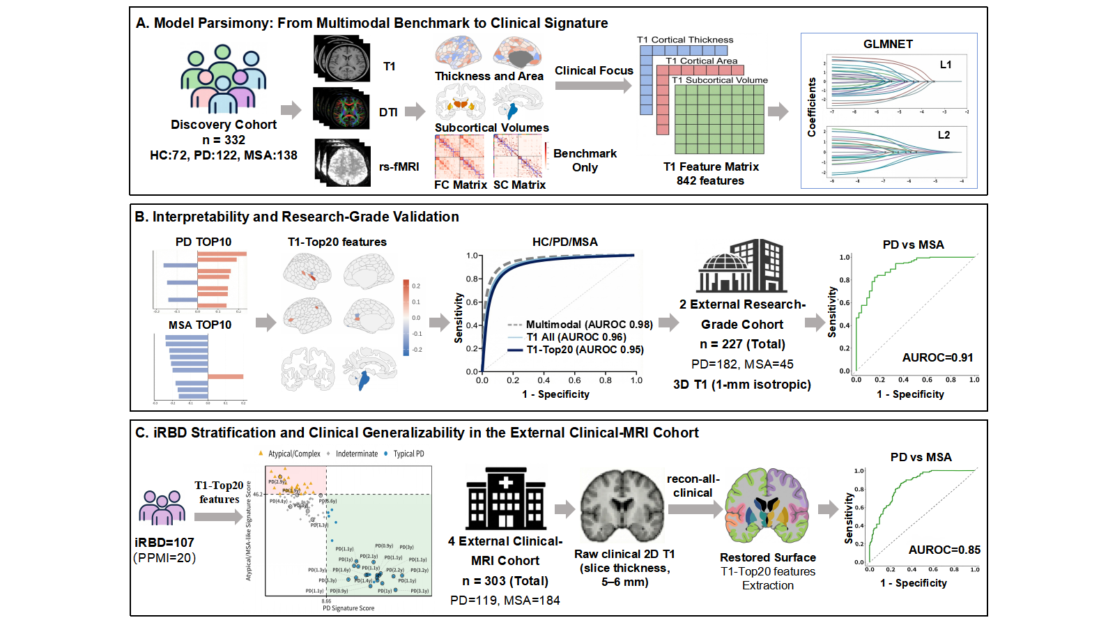
Study Workflow and Cohort Allocation
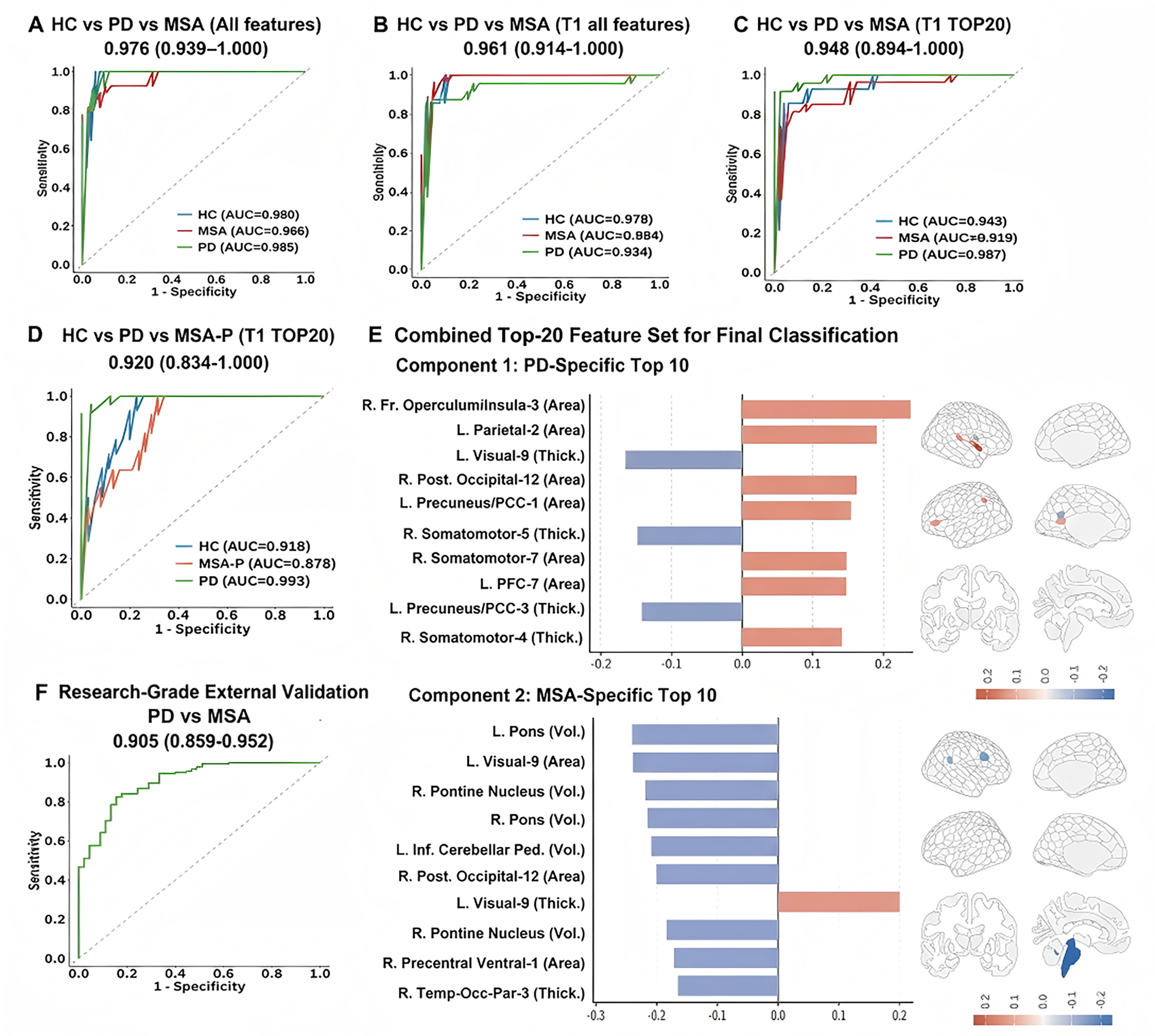
Diagnostic Performance and Interpretability
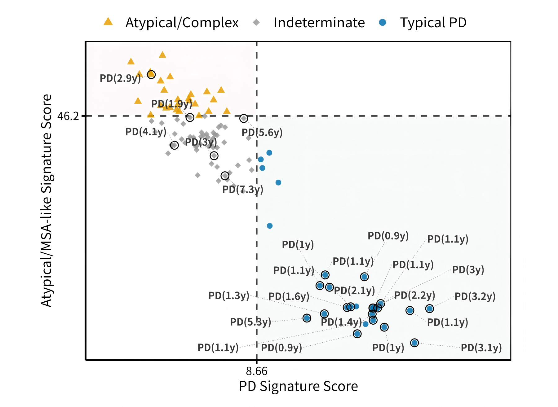
iRBD Stratification
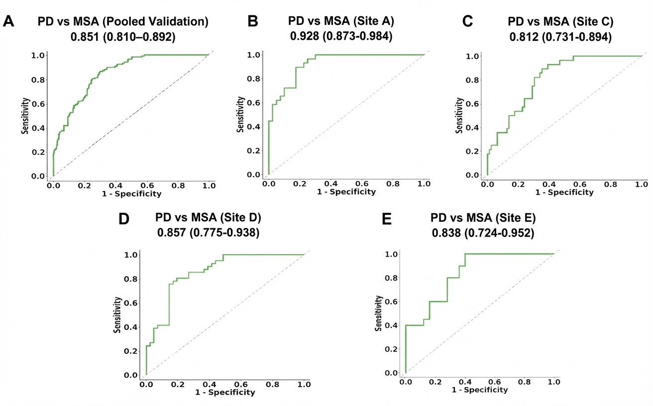
Clinical Generalizability in Clinical MRI
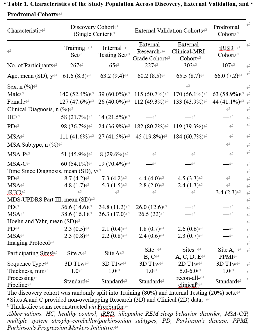
Table 1
To cite this abstract in AMA style:
LC. Zhou, CZ. Wang, AL. Du, J. Liu, J. Wang. Automated Differentiation for Early Parkinsonism Using Clinical Structural MRI: A Multicenter Study [abstract]. Mov Disord. 2026; 41 (suppl 1). https://www.mdsabstracts.org/abstract/automated-differentiation-for-early-parkinsonism-using-clinical-structural-mri-a-multicenter-study/. Accessed October 1, 2026.« Back to 2026 International Congress
MDS Abstracts - https://www.mdsabstracts.org/abstract/automated-differentiation-for-early-parkinsonism-using-clinical-structural-mri-a-multicenter-study/