
Category: Parkinson's disease: Neuroimaging
Objective: To quantitatively and qualitatively evaluate susceptibility changes in the dorsal SN (dSN), using quantitative susceptibility mapping (QSM) and susceptibility-weighted imaging (SWI) at 7Tesla (7T) MRI, to assess their potential as imaging biomarker for PD while controlling for age and sex, and to correlate the findings with clinical variables.
Background: Parkinson’s disease (PD) causes progressive motor and non-motor symptoms linked to dopaminergic neuronal loss in the substantia nigra (SN). Available imaging biomarkers sensitive to nigral pathology are limited.
Method: In this exploratory cross-sectional study, 76 healthy controls (HC) and 26 people with Parkinson’s disease (PwP) underwent 7T QSM and SWI. Focal mean susceptibility values were measured in dSN region of interest (ROI) in Sectra IDS7 [figure1], using white matter as reference. For PwP, the dSN was measured in PwP with remaining dSN hypointensity on QSM with a width of minimum 1.5 mm in the axial plane [figure2]. Grading of dSN was performed on QSM and SWI (grades 1–4). Spearman correlation analyses of QSM measurements and clinical variables were performed.
Results: The reliability of repeated focal SN measurements was good to excellent (ICC range 0.68-0.95). The focal measurements of dSN did not change with age [table 1]. Susceptibility in dSN was significantly higher in the PwP compared with HC (M=60.1 ppb vs 29.0 ppb, P< 0.0001), ANCOVA results are listed in table1. The area under the ROC curve for dSN was 0.90. The optimal cut-off value to separate PwP from HC in our material was 49.1 ppb, with a Youden’s index of 0.73 (sensitivity 83%, specificity 89%). Visual grading showed near perfect agreement between QSM and SWI (K > 0.93). A positive correlation between susceptibility in dSN and motor severity was identified (ρ = 0.60, P = 0.043). The association was not significant after correcting for multiple testing (Bonferroni/Benjamini-Hochberg). We observed that eight out of 10 (80%) of the PwP with asymmetry in dSN-grading on QSM had corresponding clinical asymmetry.
Conclusion: Our results indicate that focal susceptibility measurements in the dSN are consistently measurable in healthy controls and elevated in PwP, but due to a small number of patients findings need to be validated in larger studies.
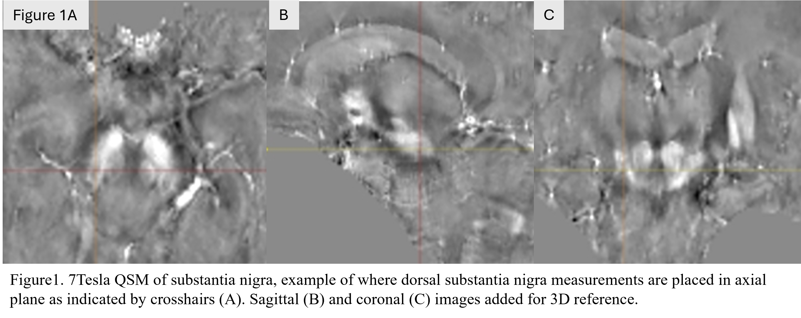
Figure 1 – 7T QSM example dSN focal ROI placement
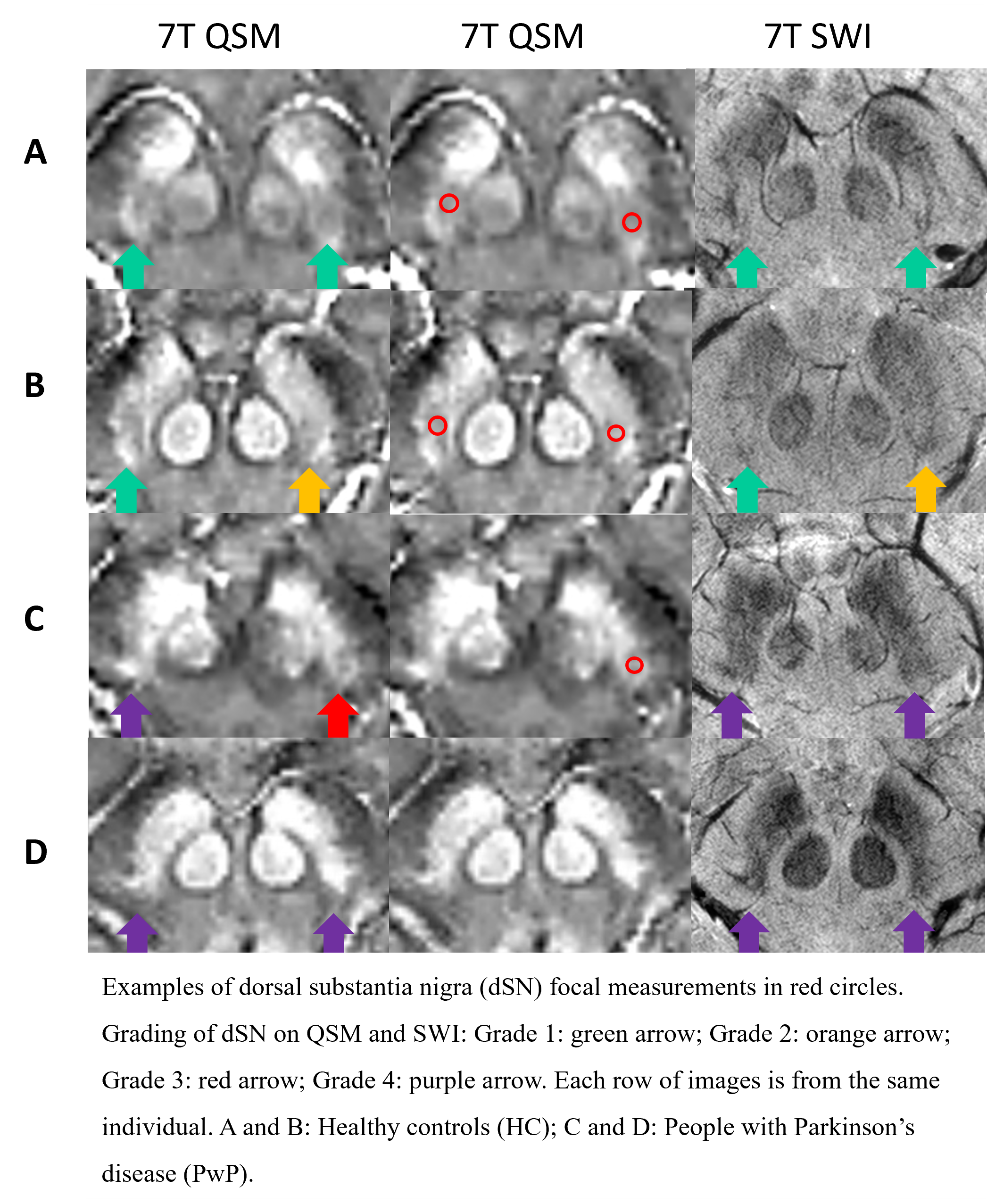
Figure 2 – Focal measurements and grading
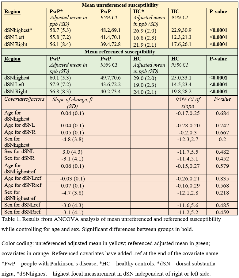
Table 1 – ANCOVA results
To cite this abstract in AMA style:
R. Unsgård, K. Stige, A. Kristoffersen, P. Goa, C. Tzoulis, E. Berntsen. 7 Tesla MRI with Quantitative Susceptibility Mapping: Quantitative focal measurements and qualitative grading of the substantia nigra as a biomarker for Parkinson’s Disease [abstract]. Mov Disord. 2026; 41 (suppl 1). https://www.mdsabstracts.org/abstract/7-tesla-mri-with-quantitative-susceptibility-mapping-quantitative-focal-measurements-and-qualitative-grading-of-the-substantia-nigra-as-a-biomarker-for-parkinsons-disease/. Accessed October 1, 2026.« Back to 2026 International Congress
MDS Abstracts - https://www.mdsabstracts.org/abstract/7-tesla-mri-with-quantitative-susceptibility-mapping-quantitative-focal-measurements-and-qualitative-grading-of-the-substantia-nigra-as-a-biomarker-for-parkinsons-disease/