
Category: Paroxysmal Movement Disorders
Objective: To present a new case of glucose transporter type 1 deficiency syndrome (GLUT1-DS) diagnosed in adulthood and to describe the associated radiological findings.
Background: Glucose transporter type 1 deficiency syndrome (GLUT1-DS) is a rare neurometabolic disorder caused by mutations in the SLC2A1 gene, resulting in impaired glucose transport across the blood-brain barrier and consequent cerebral energy deficiency. The disorder typically presents during childhood with seizures (including generalized tonic-clonic and absence seizures, among others), episodic eye-head movements, movement disorders (often triggered by fasting, fatigue or exercise) and psychomotor delay. Brain MRI findings are usually normal or show only nonspecific abnormalities, such as FLAIR hyperintensity of the U-fibers or delayed myelination.
Method: We describe a clinical case and perform a review of the relevant literature.
Results: We report a 57-year-old man previously diagnosed with cerebral palsy, who had pharmacoresistant epileptic seizures since childhood, associated with motor and intellectual disability, speech impairment and dystonic gait. He also presented paroxysmal dystonic episodes affecting both feet while walking. Brain MRI revealed marked hypointensity of the caudate nucleus and putamen (Figure 1). Genetic testing for neurodegeneration with brain iron accumulation (NBIA) disorders was negative. However, analysis of genes associated with paroxysmal dystonia identified a heterozygous pathogenic variant c.685A>T (p.Lys229Ter) in the SLC2A1 gene, resulting in a clear loss of function of the GLUT1 protein function.
Conclusion: Basal ganglia hypointensity in GLUT1-DS has been reported only once in the literature, in a series of three adult cases (1). The association between glucose metabolism and brain iron deposition has been described (3) and may reflect one of the etiopathogenic mechanisms underlying GLUT1-DS. These radiological changes may only become detectable on MRI later in adulthood. Screening for SCL2A1 variants could therefore be considered in the differential diagnosis of disorders associated with brain iron accumulation, mainly because it is a disease with a proven disease-modifying treatment (ketogenic diet).
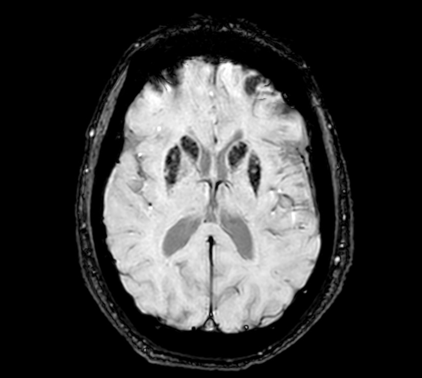
Fig 1: Image of our patient, SWI sequence at 3T.
References: 1. Van Samkar A, Leen WG, Willemsen MAAP, Verrips A. Hypointensity of the Basal Ganglia in Adults with Glucose Transporter Protein Type 1 Deficiency Syndrome: A Novel Magnetic Resonance Imaging Finding. Ann Neurol. 2020;87(1):10-11. doi:10.1002/ana.25640
2. Zhang MJ, Zhang SM, Zhang QP, et al. Clinical and genetic characteristics of glucose transporter 1 deficiency syndrome in a large cohort of Chinese patients. World J Pediatr. 2025;21(3):274-283. doi:10.1007/s12519-025-00884-9
3. Fillebeen C, Lam NH, Chow S, Botta A, Sweeney G, Pantopoulos K. Regulatory Connections between Iron and Glucose Metabolism. Int J Mol Sci. 2020;21(20):7773. Published 2020 Oct 21. doi:10.3390/ijms21207773
To cite this abstract in AMA style:
C. Jiménez Clopés, Y. Estrella Domínguez, J. Balsa Vázquez, A. Rojo Sebastián. Basal Ganglia Hypointensity Associated with Glucose Transporter Type 1 Deficiency Syndrome (GLUT1-DS) [abstract]. Mov Disord. 2026; 41 (suppl 1). https://www.mdsabstracts.org/abstract/basal-ganglia-hypointensity-associated-with-glucose-transporter-type-1-deficiency-syndrome-glut1-ds/. Accessed October 1, 2026.« Back to 2026 International Congress
MDS Abstracts - https://www.mdsabstracts.org/abstract/basal-ganglia-hypointensity-associated-with-glucose-transporter-type-1-deficiency-syndrome-glut1-ds/