
Objective: To characterize β-amyloid (Aβ) deposition across the Lewy body disease (LBD) spectrum using Pittsburgh compound B positron emission tomography (PiB-PET) and examine the influence of age, sex, and APOE ε4 genotype.
Background: Lewy body disease (LBD) represents a continuum from idiopathic REM sleep behavior disorder (iRBD) to mild cognitive impairment with Lewy bodies (MCI-LB) and dementia with Lewy bodies (DLB). Although α-synuclein pathology defines LBD, β-amyloid (Aβ) co-pathology is common, particularly in dementia. However, the stage at which amyloid accumulation emerges across the LBD spectrum remains uncertain. Clarifying its distribution may improve understanding of disease progression and inform biomarker-based therapeutic strategies.
Method: We conducted a cross-sectional study of patients who underwent 11C-Pittsburgh compound B (PiB) PET imaging between 2011 and 2025 at a dementia research center. The cohort included iRBD (n=8), MCI-LB (n=34), and probable DLB (n=42). Global cortical amyloid burden was quantified using Centiloid (CL) values derived from standardized PiB-PET processing pipelines. Aβ positivity was defined as CL>20. Group differences were assessed using appropriate statistical tests, and multivariable regression examined associations between CL values and age, sex, APOE ε4 status, and cognition.
Results: In this cohort of 82 patients, global cortical amyloid burden differed across groups (p<0.001). Mean CL values were highest in DLB (47.8±39.3), intermediate in MCI-LB (19.9±32.9), and minimal in iRBD (1.65±7.52). Aβ positivity increased along the spectrum: 67% in DLB, 32% in MCI-LB, and none in iRBD. Higher CL values were associated with poorer cognition, showing negative correlations with MMSE (r=−0.306, p=0.005) and HDS-R (r=−0.335, p=0.002). APOE ε4 carriers had higher CL values in both MCI-LB (p=0.003) and DLB (p=0.015). Age was positively associated with CL values, whereas sex showed no association.
Conclusion: Amyloid deposition increases progressively across the LBD spectrum, with minimal accumulation in iRBD, intermediate levels in MCI-LB, and the highest burden in DLB. These findings suggest cortical amyloid deposition emerges mainly from the prodromal stage rather than the earliest stage of LBD. Amyloid-positive MCI-LB may represent a promising stage for biomarker-guided disease-modifying trials targeting amyloid pathology in Lewy body diseases.
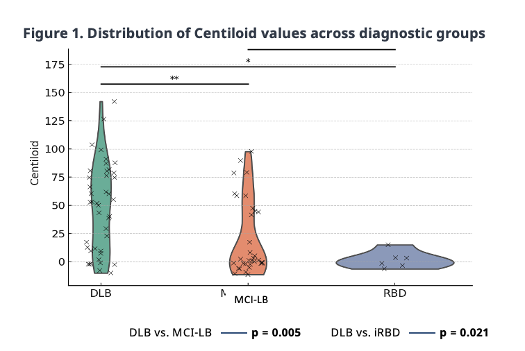
Fig1. Distribution of centiloid values
References: Kobayashi R, Hayashi H, Kawakatsu S, Okamura N, Yoshioka M, Otani K. Assessment of amyloid deposition in patients with probable REM sleep behavior disorder as a prodromal symptom of dementia with Lewy bodies using PiB-PET. Front Neurol. 2019;10:671.
Gomperts SN, Rentz DM, Moran E, et al. Imaging amyloid deposition in Lewy body diseases. Neurology. 2008;71(12):903-910.
Nedelska Z, Schwarz CG, Lesnick TG, et al. Association of longitudinal β-amyloid accumulation determined by positron emission tomography with clinical and cognitive decline in adults with probable Lewy body dementia. JAMA Netw Open. 2019; 2(12):e1916439.
To cite this abstract in AMA style:
M. Yoshita, S. Tsuji, K. Yokoyama. Emergence of Cortical Amyloid from the Prodromal Stage of the Lewy Body Disease Spectrum: Evidence from a PiB-PET Study [abstract]. Mov Disord. 2026; 41 (suppl 1). https://www.mdsabstracts.org/abstract/emergence-of-cortical-amyloid-from-the-prodromal-stage-of-the-lewy-body-disease-spectrum-evidence-from-a-pib-pet-study/. Accessed October 1, 2026.« Back to 2026 International Congress
MDS Abstracts - https://www.mdsabstracts.org/abstract/emergence-of-cortical-amyloid-from-the-prodromal-stage-of-the-lewy-body-disease-spectrum-evidence-from-a-pib-pet-study/