
Category: Parkinson's disease: Neuroimaging
Objective: To study the association between multimodal non-invasive MRI glymphatic system proxies and global and deep gray matter (GM) degeneration in patients with Parkinson’s disease (PD).
Background: Glymphatic dysfunction has been hypothesized to impair pathological protein clearance and to exacerbate downstream pathological mechanisms in synucleinopathies, ultimately contributing to neuronal loss [1]. However, only two previous studies have reported associations between the Diffusion-Tensor Imaging Along the Perivascular Space (DTI-ALPS) index, a widely used glymphatic proxy, and cortical GM volume [2,3], and only one study has examined its relationship with subcortical nuclei volume, yielding no significant association [2]. Notably, nowadays additional magnetic resonance imaging (MRI) glymphatic proxies have been proposed to complement the DTI-ALPS index [4], providing a more comprehensive picture for understanding the glymphatic functioning.
Method: Fifty-two patients with PD and 35 controls underwent an MRI assessment. FLAIR, T1- and diffusion-weighted imaging sequences were used to calculate DTI-ALPS indices, free-water content in white matter, perivascular spaces count and volume, and mean choroid plexus (CP) volume. Global atrophy and deep GM measures were introduced in further analysis as volume/TIV. Between-group differences in glymphatic proxies and its relationship with atrophy measures were assessed adjusting by age. The significant threshold was set at p<0.05.
Results: Patients with PD showed lower right, left and bilateral DTI-ALPS indices and higher CP volume compared with controls [Figure 1]. Patients with PD also hadlower total and cortical GM volume and lower amygdala and putamen volume [Table 1]. A positive correlation was found between bilateral DTI-ALPS index and amygdala and putamen volumes in patients with PD [Figure 2]. No other correlations were found with DTI-ALPS indices or CP volume.
Conclusion: Our findings provide novel evidence that glymphatic waste‑clearance functioning is impaired in PD and is associated with neurodegeneration in disease‑relevant nuclei such as the putamen and amygdala. By integrating MRI‑based glymphatic proxies beyond the traditional DTI‑ALPS index, this study offers a broader and more comprehensive framework for understanding glymphatic system involvement in PD.
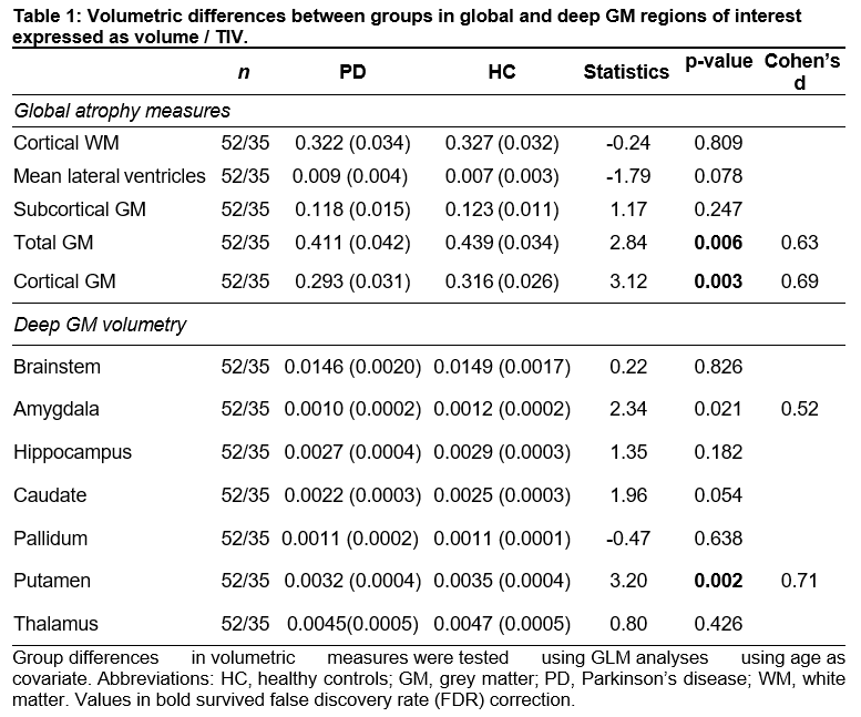
Table 1.
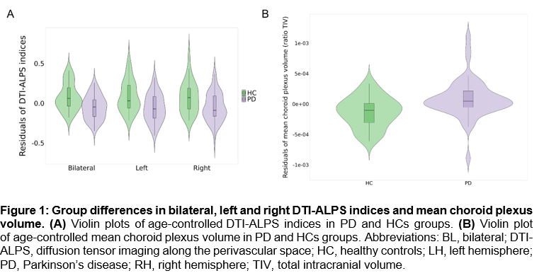
Figure 1.
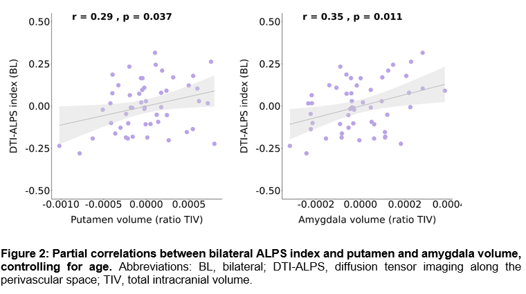
Figure 2.
References: [1] Sun, Y., Lv, Q.-K., Liu, J.-Y., Wang, F. & Liu, C.-F. New perspectives on the glymphatic system and the relationship between glymphatic system and neurodegenerative diseases. Neurobiol. Dis. 205, 106791 (2025).
[2] Zhu, Z. et al. Glymphatic dysfunction is related to motor disability fully mediated by gray matter degeneration in early-stage Parkinson’s disease. Neurobiol. Dis. 213, 107001 (2025).
[3] Zhao, Y. Et al. Glymphatic dysfunction exacerbates cognitive decline by triggering cortical degeneration in Parkinson’s disease: evidence from diffusion-tensor MRI. Brain Commun. 7, fcaf029 (2024).
[4] Zhou, X. et al. Glymphatic, Structural, and Cognitive Changes During Breast Cancer Chemotherapy: A Longitudinal MRI Study. Hum. Brain Mapp. 46, e70334 (2025).
To cite this abstract in AMA style:
C. Garcia-Vicente, J. Pardo, C. Martín-Barceló, I. Roura, C. Falcon, R. Sala-Llonch, A. Campabadal, C. Uribe, N. Bargalló, O. Brengaret, C. Brenlla, C. Painous, A. Cámara, M. Buongiorno, M. Martí, Y. Compta, C. Junqué, B. Segura. Glymphatic dysfunction in patients with Parkinson’s disease is associated with regional amygdala and putamen degeneration [abstract]. Mov Disord. 2026; 41 (suppl 1). https://www.mdsabstracts.org/abstract/glymphatic-dysfunction-in-patients-with-parkinsons-disease-is-associated-with-regional-amygdala-and-putamen-degeneration/. Accessed October 1, 2026.« Back to 2026 International Congress
MDS Abstracts - https://www.mdsabstracts.org/abstract/glymphatic-dysfunction-in-patients-with-parkinsons-disease-is-associated-with-regional-amygdala-and-putamen-degeneration/