
Category: Parkinson's disease: Neuroimaging
Objective: To test whether GBA1 mutations are associated with impaired cortical compensation in Parkinson’s disease (PD) using a unique 2×2 design to parse the effect of gene and disease.
Background: GBA1, encoding glucocerebrosidase, harbors the strongest known genetic risk variants for PD that are associated with a more severe phenotype. Recently, we have demonstrated that motor impairment in PD is characterized by loss of compensatory function in the parieto-premotor cortex. Given evidence for greater cortical pathology in GBA1-variant carriers in PD (PDGBA1+), cortical compensation may be disproportionally impaired.
Method: 90 participants (controls: GBA1+ n=24, GBA1− n=24; PD: GBA1+ n=16, GBA1− n=26; mean PD duration 5.59 years) from the Luxembourg Parkinson’s Study1 (LuxPark) underwent 3T functional MRI while performing an action selection task designed to elicit PD-related compensatory activity. Diffusion MRI was additionally used to investigate underlying structural deficits in the substantia nigra and cortex. GBA1 status were detected by long-read and Sanger sequencing. Variant pathogenicity was classified using the GBA1-PD browser. Linear mixed-effects modeling was used to analyze effects of disease (PD vs. Control) and genetic status (GBA1- vs. GBA1+) on behavioral performance and brain measurements. Findings were validated in the Personalized Parkinson Project2 (PPP; 286 PDGBA1− and 49 PDGBA1+, mean PD duration 2.76 years).
Results: In LuxPark, PDGBA1+ made more errors on the task (diagnosis × GBA1 interaction, p=0.01) and tended to have greater motor impairment (MDS-UPDRS part III, p=0.09) than PDGBA-. PDGBA+ also had lower parieto-premotor selection-related activity, suggesting reduced compensation (diagnosis × GBA1 interaction, p<0.001, which correlated with higher clinical bradykinesia/rigidity [figure1]. Nigral free water was increased in PD (p<0.001) but did not differ by GBA1 status. Parietal mean diffusivity was higher in PDGBA1+ (p=0.021, uncorrected), which was associated with higher bradykinesia/rigidity [figure2]. In PPP, GBA1 pathogenicity was associated with reduced selection-related activity (p=0.012). There were no further GBA1-related effects on clinical or MRI measurements.
Conclusion: Our findings suggest that GBA1 variants impair cortical compensation, potentially by exacerbating microstructural cortical damage. This effect depends on variant pathogenicity and emerges with advancing disease.
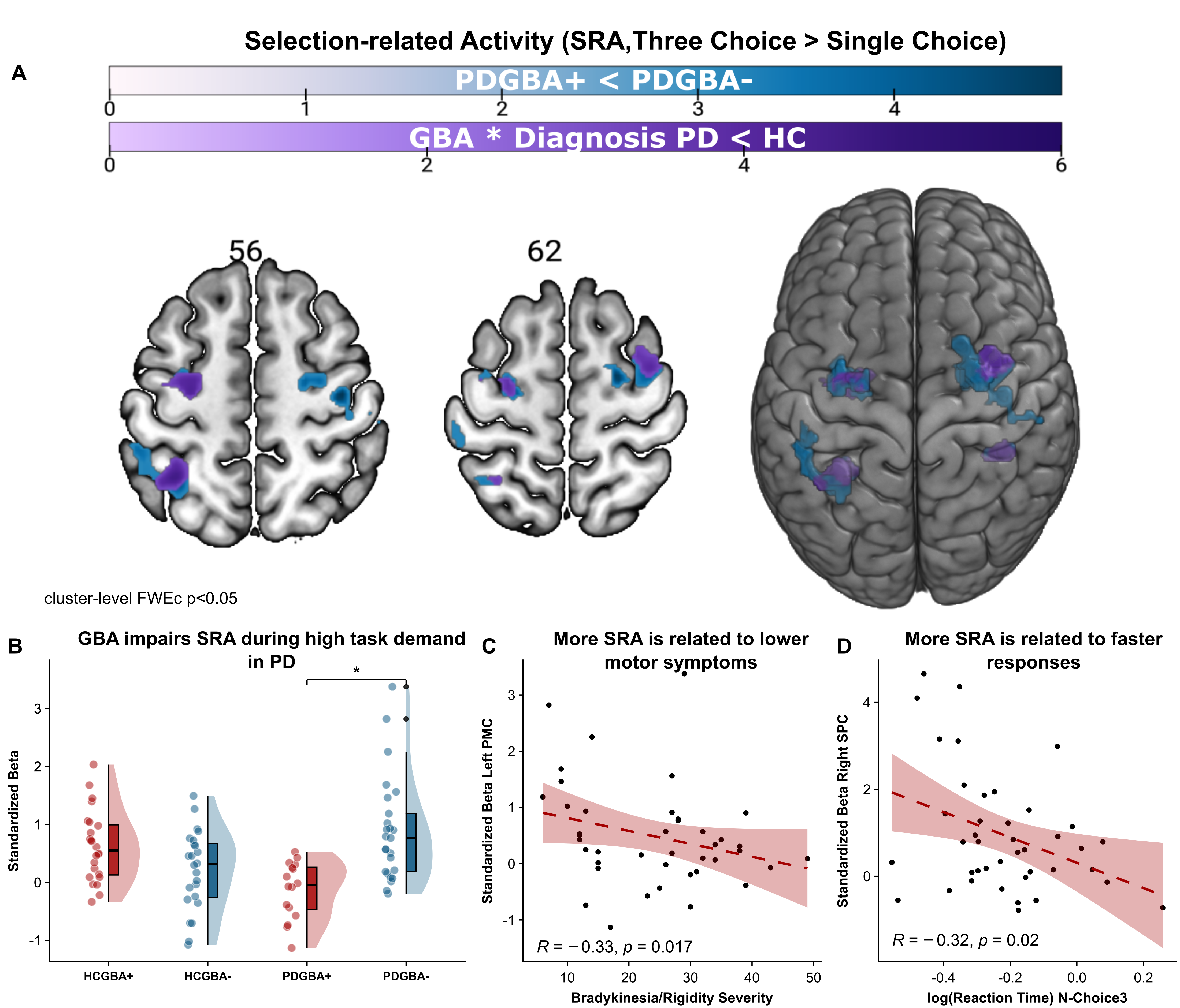
Figure1: GBA1 impairs cortical compensation in PD
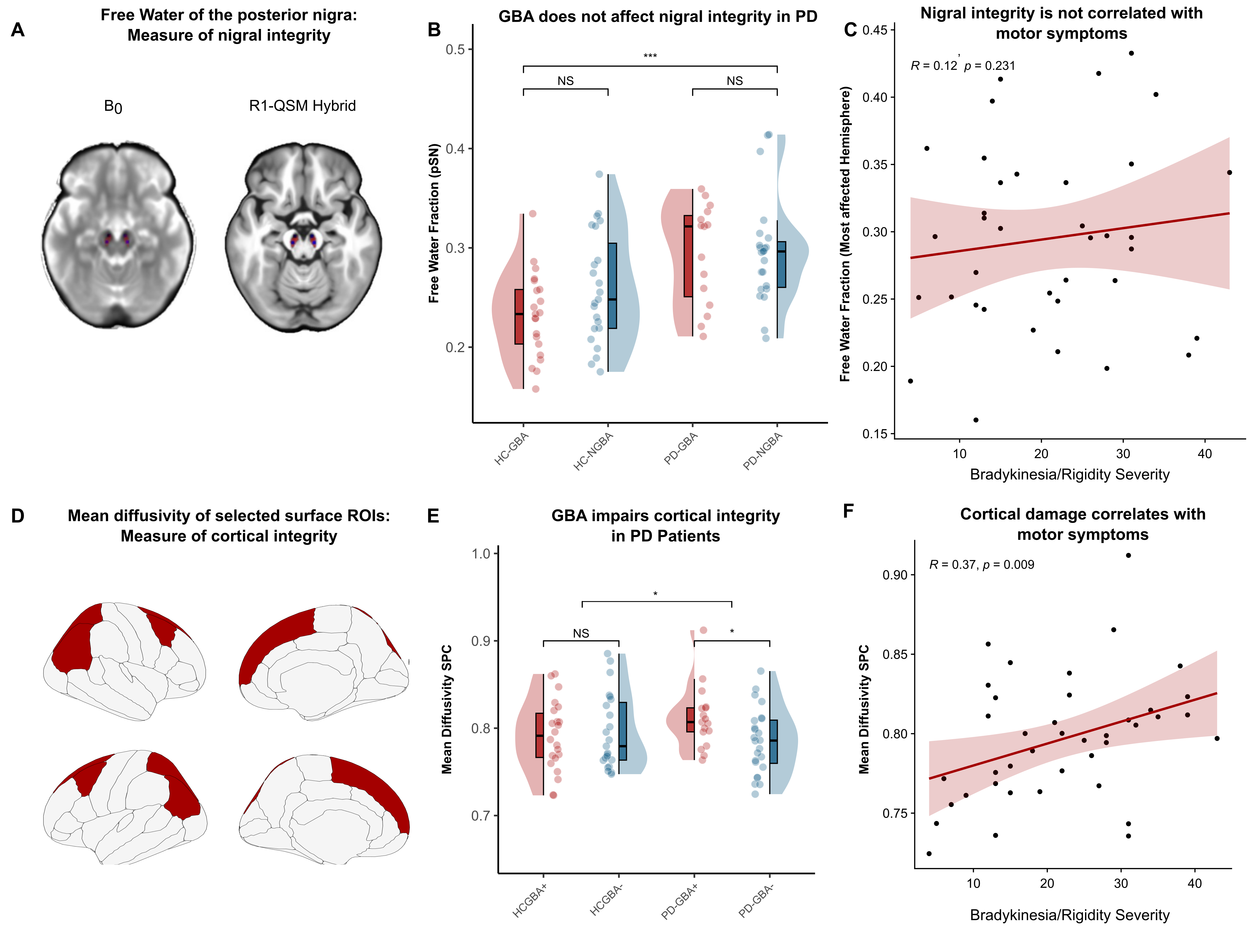
Figure 2: GBA1 impairs cortical integrity in PD
References: 1. Pavelka L, Rawal R, Ghosh S, et al. Luxembourg Parkinson’s study -comprehensive baseline analysis of Parkinson’s disease and atypical parkinsonism. Front Neurol. 2023;14:1330321. doi:10.3389/fneur.2023.1330321
2. Bloem BR, Marks WJ, Silva De Lima AL, et al. The Personalized Parkinson Project: examining disease progression through broad biomarkers in early Parkinson’s disease. BMC Neurol. 2019;19(1):160. doi:10.1186/s12883-019-1394-3
To cite this abstract in AMA style:
K. Steidel, M. Johansson, B. Bloem, L. Pavelka, T. Lüth, C. Pauly, E. Soboleva, S. Jónsdóttir, C. Much, S. Schaake, L. Vilas Boas, E. Thiry, G. Zelimkhanov, N. Brüggemann, C. Klein, J. Trinh, R. Krüger, R. Helmich. Impaired Cortical Compensation in GBA1-Associated Parkinson’s Disease [abstract]. Mov Disord. 2026; 41 (suppl 1). https://www.mdsabstracts.org/abstract/impaired-cortical-compensation-in-gba1-associated-parkinsons-disease/. Accessed October 1, 2026.« Back to 2026 International Congress
MDS Abstracts - https://www.mdsabstracts.org/abstract/impaired-cortical-compensation-in-gba1-associated-parkinsons-disease/