
Category: Parkinson's disease: Neuroimaging
Objective: To investigate the distribution and potential influence of tau pathology in Parkinson’s disease (PD) patients with and without prodromal REM sleep behavior disorder (pRBD), using 18F-Florzolotau positron emission tomography (PET) imaging.To investigate the distribution and potential influence of tau pathology in Parkinson’s disease (PD) patients with and without prodromal REM sleep behavior disorder (pRBD), using 18F-Florzolotau positron emission tomography (PET) imaging.
Background: Recent models propose that PD can be stratified into the body-first and brain-first subtypes based on the origin of α-synuclein pathology. Importantly, this hypothesis suggests pRBD as a key marker to distinguish these two phenotypes(1). Meanwhile, several studies regarding PD pathogenesis have corroborated distinct patterns of tau accumulation. While tau co-pathology may play a critical role in α-synucleinopathy progression(2), whether tau pathology diverges across these putative PD subtypes has not been delineated.
Method: We recruited twenty-six PD patients and classified them into two groups based on the REM Sleep Behavior Disorder Screening Questionnaire (RBDSQ). Sixteen patients who scored ≤3 were classified as putative brain-first subtype (PDpRBD-), and ten patients who scored ≥6 as putative body-first subtype (PDpRBD+). All participants underwent clinical assessments and 18F-Florzolotau PET imaging. Intergroup differences and clinical correlations were assessed using voxel-based analyses.
Results: Voxel-based 18F-Florzolotau uptake was significantly higher in PDpRBD+ patients compared to the PDpRBD- group, particularly in the right precuneus and left posterior cingulate regions. Notably, in the putative body-first cohort, higher tau tracer binding in the right limbic system, specifically the right putamen (R=-0.831, P=0.003) and right insula (R=-0.790, P=0.007), was inversely correlated with cognitive performance, as measured by the Mini-Mental State Examination (MMSE) scores, after adjusting for age, gender, and education.
Conclusion: Our study demonstrated distinct patterns of tau tracer uptake between PDpRBD+ and PDpRBD- patients. Notably, the putative body-first phenotype exhibited a more extensive tau burden that correlated with cognitive impairment. These results provide in vivo imaging evidence on the tau pathology divergence of hypothesized PD subtypes.
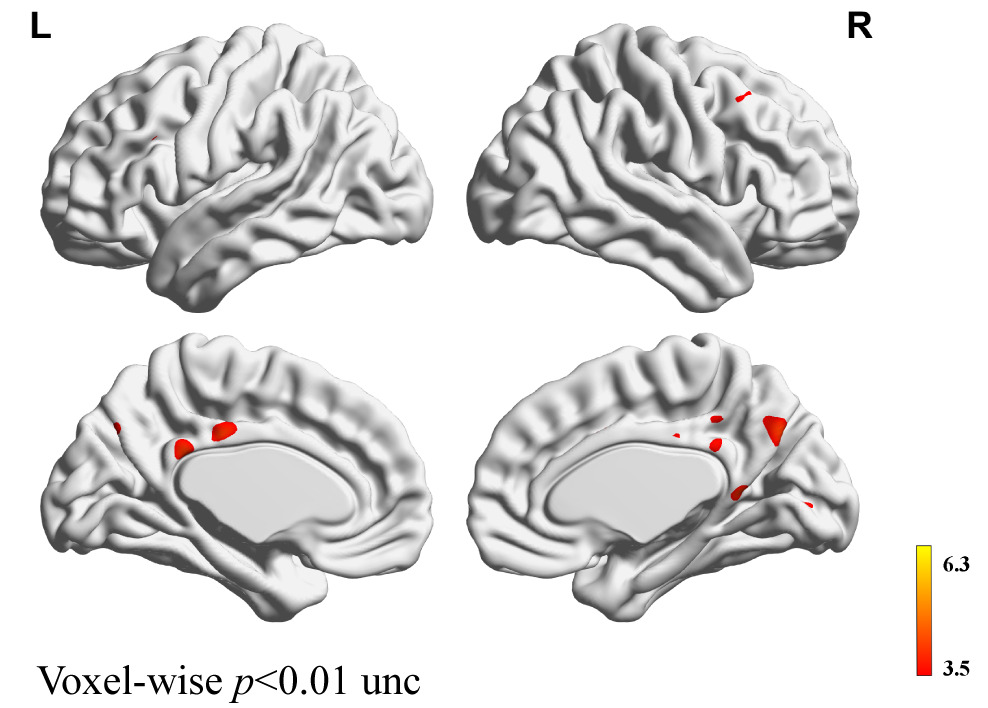
Voxel-wise Tau PET comparison: PDpRBD+ vs PDpRBD-
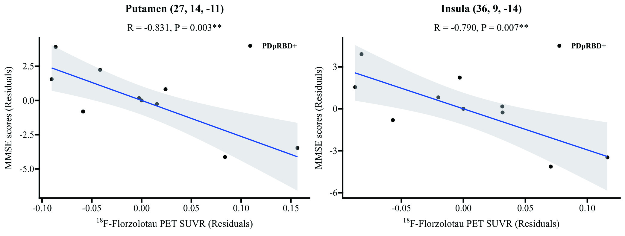
Tau PET uptake and MMSE Correlation in PDpRBD+
References: 1. McKenna D, Peever J. Degeneration of rapid eye movement sleep circuitry underlies rapid eye movement sleep behavior disorder. Mov Disord 2017;32(5):636–644. https://doi.org/10.1002/mds.27003
2. Vermilyea SC, Christensen A, Meints J, et al. Loss of tau expression attenuates neurodegeneration associated with α-synucleinopathy. Transl Neurodegener 2022;11(1):34. https://doi.org/10.1186/s40035-022-00309-x
To cite this abstract in AMA style:
C. Xu, Y. Tang, J. Wang. In Vivo 18F-Florzolotau Tau PET Imaging in Parkinson’s Disease with and without Probable REM Sleep Behavior Disorder [abstract]. Mov Disord. 2026; 41 (suppl 1). https://www.mdsabstracts.org/abstract/in-vivo-18f-florzolotau-tau-pet-imaging-in-parkinsons-disease-with-and-without-probable-rem-sleep-behavior-disorder/. Accessed October 1, 2026.« Back to 2026 International Congress
MDS Abstracts - https://www.mdsabstracts.org/abstract/in-vivo-18f-florzolotau-tau-pet-imaging-in-parkinsons-disease-with-and-without-probable-rem-sleep-behavior-disorder/