
Objective: To synthesize current experimental and translational evidence supporting the hypothesis that micro- and nanoplastics (MNPs) may contribute to Parkinson’s disease (PD) pathogenesis through neuroinflammatory and oxidative stress–related mechanisms.
Background: Parkinson’s disease (PD) is a progressive neurodegenerative disorder resulting from interactions between genetic susceptibility and environmental exposures. Micro- and nanoplastics (MNPs) are ubiquitous environmental pollutants with emerging evidence of neurotoxic effects, raising concerns about their potential contribution to neurodegenerative processes.
Method: A narrative review of peer-reviewed experimental, toxicological, and human biomonitoring studies was conducted. The analysis focused on evidence related to systemic distribution of MNPs, central nervous system accessibility, microglial activation, oxidative stress, mitochondrial dysfunction, and alpha-synuclein–related pathways relevant to PD.
Results: MNPs have been detected in human blood, lungs, placenta, cerebrospinal fluid, and brain tissue [1,2]. Experimental studies indicate that MNPs can cross biological barriers and access the brain via olfactory pathways or disruption of the blood–brain barrier. In vitro and in vivo models demonstrate that MNP exposure induces oxidative stress, mitochondrial dysfunction, apoptosis, and microglial activation [3,4]. These mechanisms overlap with established pathogenic processes in PD, including neuroinflammation and alpha-synuclein–mediated neurotoxicity [5]. However, evidence linking MNP exposure to PD in humans remains indirect.
Conclusion: Current experimental and translational data support the biological plausibility of micro- and nanoplastics as environmental contributors to Parkinson’s disease–related neurodegeneration. Although direct clinical evidence is limited, these findings highlight the need to consider MNPs as potentially modifiable environmental risk factors. Further studies incorporating standardized exposure assessment, long-term models, and human biomonitoring are required to clarify their role in PD pathogenesis.
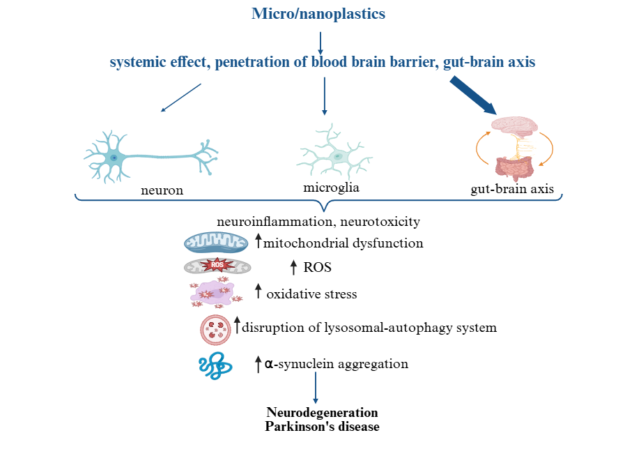
Micro- and nanoplastics in Parkinson’s disease
References: 1. Leslie HA, van Velzen MJM, Brandsma SH, Vethaak AD, Garcia-Vallejo JJ, Lamoree MH. Discovery and quantification of plastic particle pollution in human blood. Environ Int. 2022;163:107199.
2. Horvatits T, Twerenbold R, Meier F, et al. Microplastics detected in human cerebrospinal fluid and brain tissue. Sci Total Environ. 2024;912:167958.
3. Prüst M, Meijer J, Westerink RHS. The plastic brain: neurotoxicity of micro- and nanoplastics. Part Fibre Toxicol. 2020;17(1):24.
4. Luo H, Xiang Y, He D, Li Y, Zhao Y, Wang S, et al. Polystyrene microplastics induce neurotoxicity through oxidative stress and apoptosis in Caenorhabditis elegans. Environ Toxicol Pharmacol. 2019;69:37–43.
5. Zhang W, Wang T, Pei Z, Miller DS, Wu X, Block ML, et al. Aggregated alpha-synuclein activates microglia: a process leading to disease progression in Parkinson’s disease. FASEB J. 2005;19(6):533–42.
To cite this abstract in AMA style:
A. Aralbayeva, Z. Akhmetzhanova, A. Baidarbekova. Micro- and Nanoplastics in Parkinson’s Disease Pathogenesis An Emerging Environmental Risk Factor [abstract]. Mov Disord. 2026; 41 (suppl 1). https://www.mdsabstracts.org/abstract/micro-and-nanoplastics-in-parkinsons-disease-pathogenesis-an-emerging-environmental-risk-factor/. Accessed October 1, 2026.« Back to 2026 International Congress
MDS Abstracts - https://www.mdsabstracts.org/abstract/micro-and-nanoplastics-in-parkinsons-disease-pathogenesis-an-emerging-environmental-risk-factor/