
Objective: To compare regional brain volumes using voxel-based morphometry (VBM) and brain network patterns during imagined writing (IW) and actual writing (WT) using task based functional magnetic resonance imaging (fMRI) in healthy volunteers (HV) and writer’s cramp (WC).
Background: This study investigates task-specific dystonia by examining structural volumes and functional connectivity of the inferior parietal lobule during active and imagined writing. The research specifically focuses on how this region interacts with key motor network nodes to better understand the aberrant neural circuitry underlying Writer’s Cramp.
Method: We recruited 15 sWC (mean age: 32.2 ± 10.35 yrs) , and 9 HV (mean age: 29.66 ± 3.39 yrs) and acquired 3T MRl. TR/TE = 8.1/3.7 ms. fMRI data were acquired during a block-design task- 12 active blocks [IW and WT] of 40s alternating with 10 rest blocks of 20s (TR/TE = 1000/25 ms; 710 dynamics).
fMRI connectivity results were evaluated using Fisher-transformed correlations with voxel-level pFDR < 0.01 and cluster-level pFDR < 0.05.
Results: The VBM analysis revealed no significant structural differences between WC and HV at pFDR < 0.05. However, total intracranial volume (TIV) was found to be higher in WC (Figure 1, Figure 2).
fMRI revealed distinct activation patterns between HV and WC during both IW and WT tasks. In both groups, the superior parietal lobule (SPL) and supplementary motor area (SMA) remained consistently activated.
WC exhibited notable network alterations: During WT , inhibition of the posterior cingulate gyrus (pCG) within the default mode network (DMN) was observed, along with increased activation of the supramarginal gyrus (SMG). During IW, bilateral inhibition of the angular gyrus (AG) was detected, while the superior temporal gyrus showed enhanced connectivity with the precentral gyrus. During WT, inhibition of the left AG and hippocampus was accompanied by increased activation of the cerebellar and sensorimotor networks (Figure 3).
Conclusion: WC was not associated with significant regional structural brain changes as measured by VBM. Reduced default mode network activity along with increased engagement of sensorimotor and cerebellar networks, suggests compensatory mechanisms and altered network integration that may contribute to impaired motor control in WC.
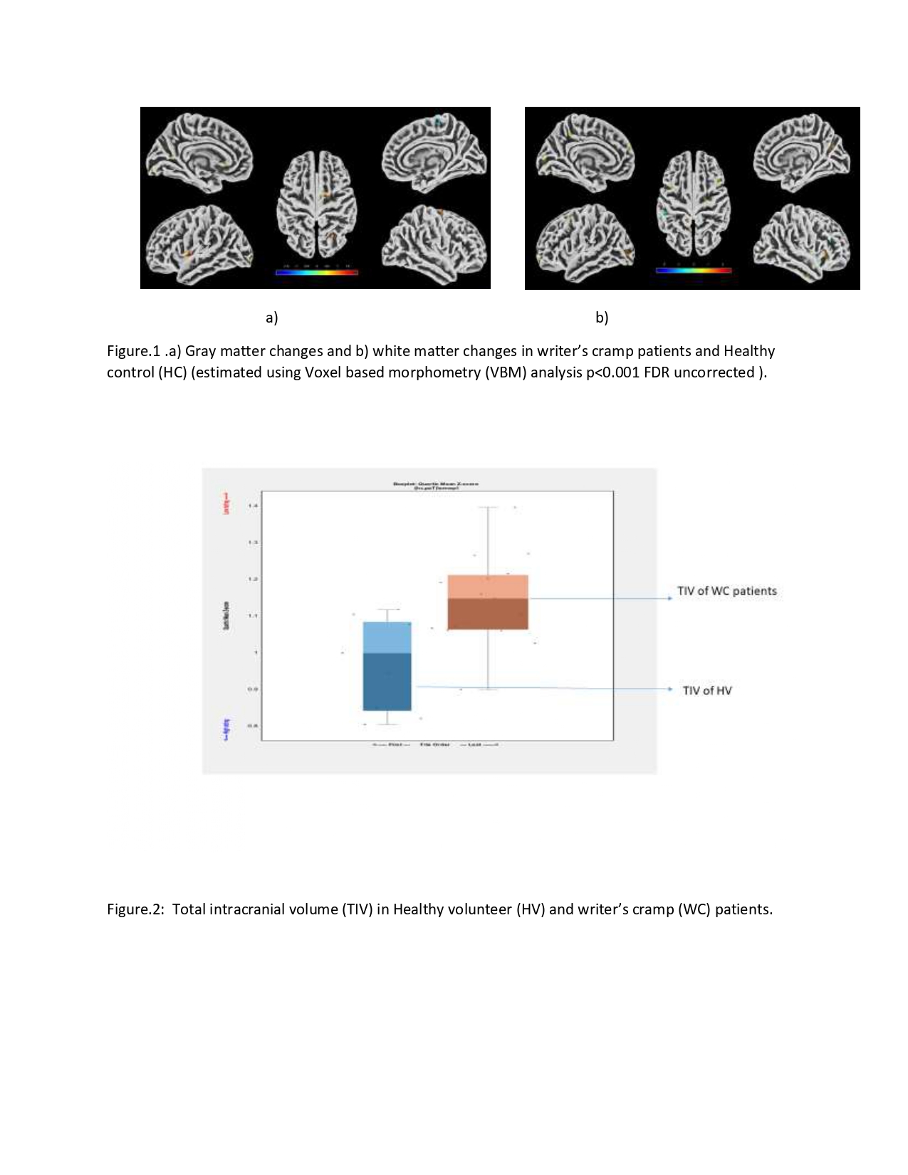
Figure 1 and Figure 2
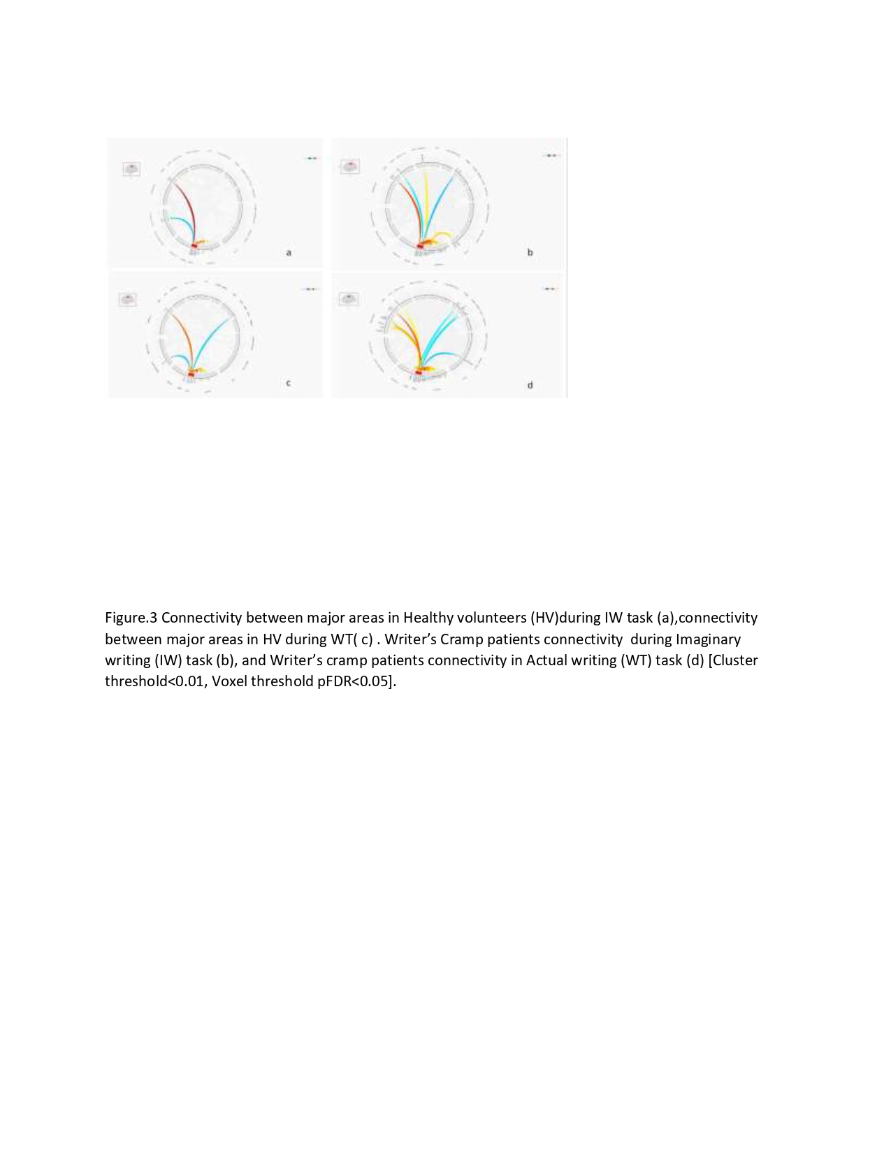
Figure 3
References: 1. Baumann A, Gless CA, Knutzen A, Granert O, Tödt I, Schmidt J, Gerke T, Wolff S, Marquardt C, Witt K, Zeuner KE. Kinaesthetic motor imagery in writer’s cramp dystonia reveals writing specific abnormalities in the occipital lobe. Neuroscience. 2025 Sep 27;589:221-229. doi: 10.1016/j.neuroscience.2025.09.042. Epub ahead of print. PMID: 41022349.
2. https://www.nitrc.org/projects/conn
To cite this abstract in AMA style:
D. Biswas, P. Pankaj, P. Bhat, D. M.R, A. Upadhyay, A. Srivastava, N. Kumar, S. Kumaran, R. Rajan. Regional Brain Volumes and Network Alterations During Task-Based fMRI in Writer’s Cramp: A Comparison with Healthy Controls [abstract]. Mov Disord. 2026; 41 (suppl 1). https://www.mdsabstracts.org/abstract/regional-brain-volumes-and-network-alterations-during-task-based-fmri-in-writers-cramp-a-comparison-with-healthy-controls/. Accessed October 1, 2026.« Back to 2026 International Congress
MDS Abstracts - https://www.mdsabstracts.org/abstract/regional-brain-volumes-and-network-alterations-during-task-based-fmri-in-writers-cramp-a-comparison-with-healthy-controls/