
Category: Parkinson's disease: Neuroimaging
Objective: To investigate the spatiotemporal dynamics of multimodal biomarkers and their role in early prediction of cognitive decline.
Background: Cognitive impairment is a major source of disability in Parkinson’s disease (PD), yet reliable predictive biomarkers remain limited. Glial fibrillary acidic protein (GFAP) and neurofilament light chain (NfL) reflect neuroinflammation and axonal degeneration, and the diffusion MRI-derived free water (FW) and intracellular volume fraction (ICVF) reflect extracellular and axonal structural changes, respectively.
Method: We analyzed 99 individuals with clinically established or probable PD who underwent plasma sampling and diffusion MRI at baseline and 1–3-year follow-up. FW and ICVF were quantified in the nucleus basalis of Meynert (NBM) and white-matter tracts linked to cognitive impairment. Associations between baseline plasma biomarkers and longitudinal MRI changes were assessed using covariate-adjusted regression and tract-based spatial statistics. One-year cognitive decline was predicted using gradient boosting, and feature importance was evaluated. To infer the temporal ordering, we modeled longitudinal trajectories using hierarchical Bayesian modeling.
Results: Higher baseline GFAP levels were associated with greater increases in FW in those with normal cognition, strongest in the anterior limb of the internal capsule (β=0.45, q=0.019). In contrast, higher baseline NfL levels were associated with greater decreases in ICVF in those with cognitive impairment, strongest in the superior longitudinal fasciculus (β=−0.63, q=0.029) [table1]. Tract-based spatial statistics confirmed these distinct patterns. Machine learning identified FW and GFAP as the strongest contributors to 1-year cognitive prediction, with the NBM and anterior limb of the internal capsule providing the highest importance. Hierarchical sigmoid modeling revealed a temporal sequence that began with free water increase in the NBM, was followed by changes in other biomarkers, and culminated in NfL elevation [figure1].
Conclusion: Plasma biomarkers and diffusion MRI metrics capture complementary inflammatory and degenerative processes in PD, supporting a NBM-centered model in which early neuroinflammatory changes precede subsequent white-matter degeneration and cognitive decline, supporting early risk stratification and therapeutic intervention.
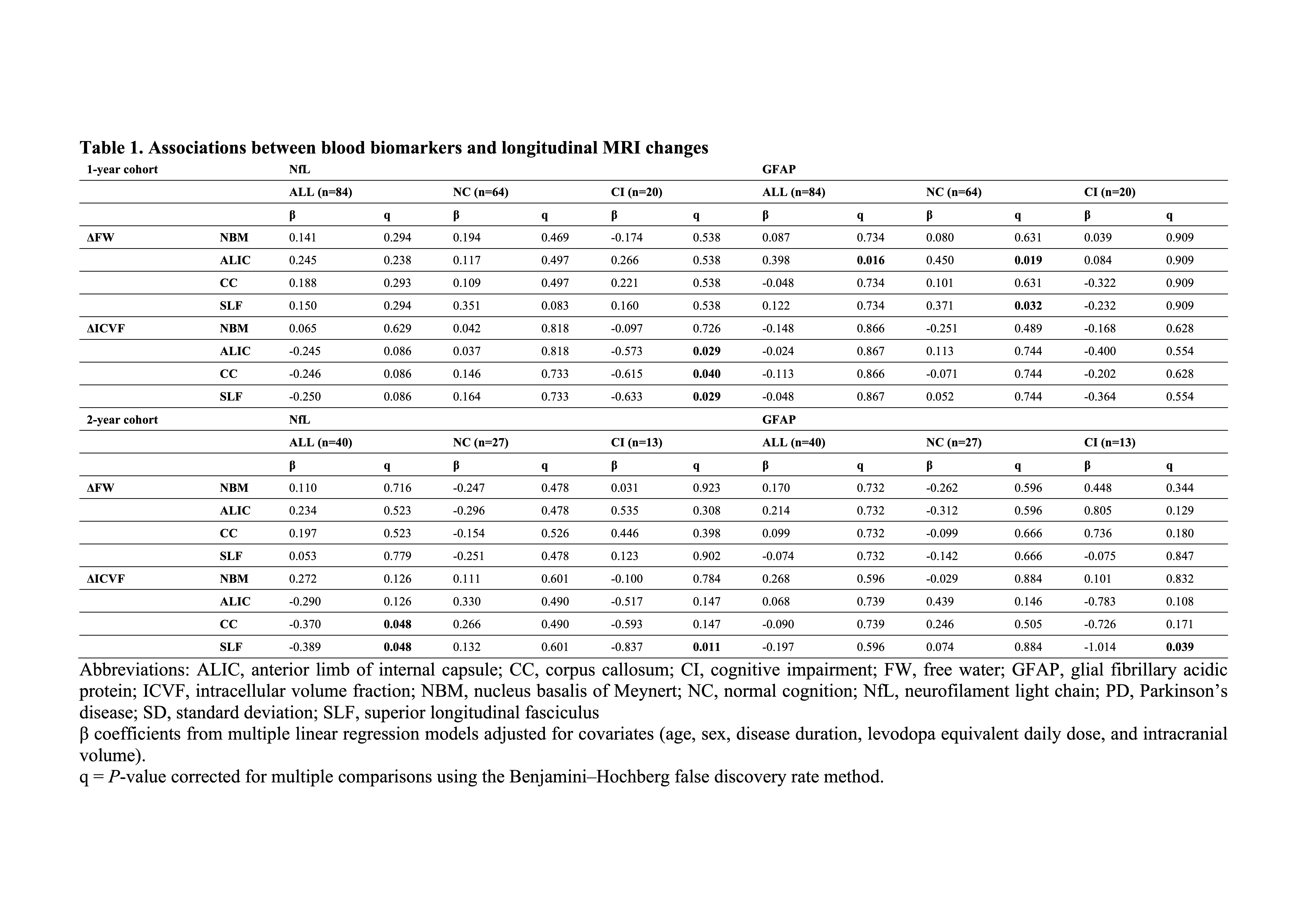
Table 1
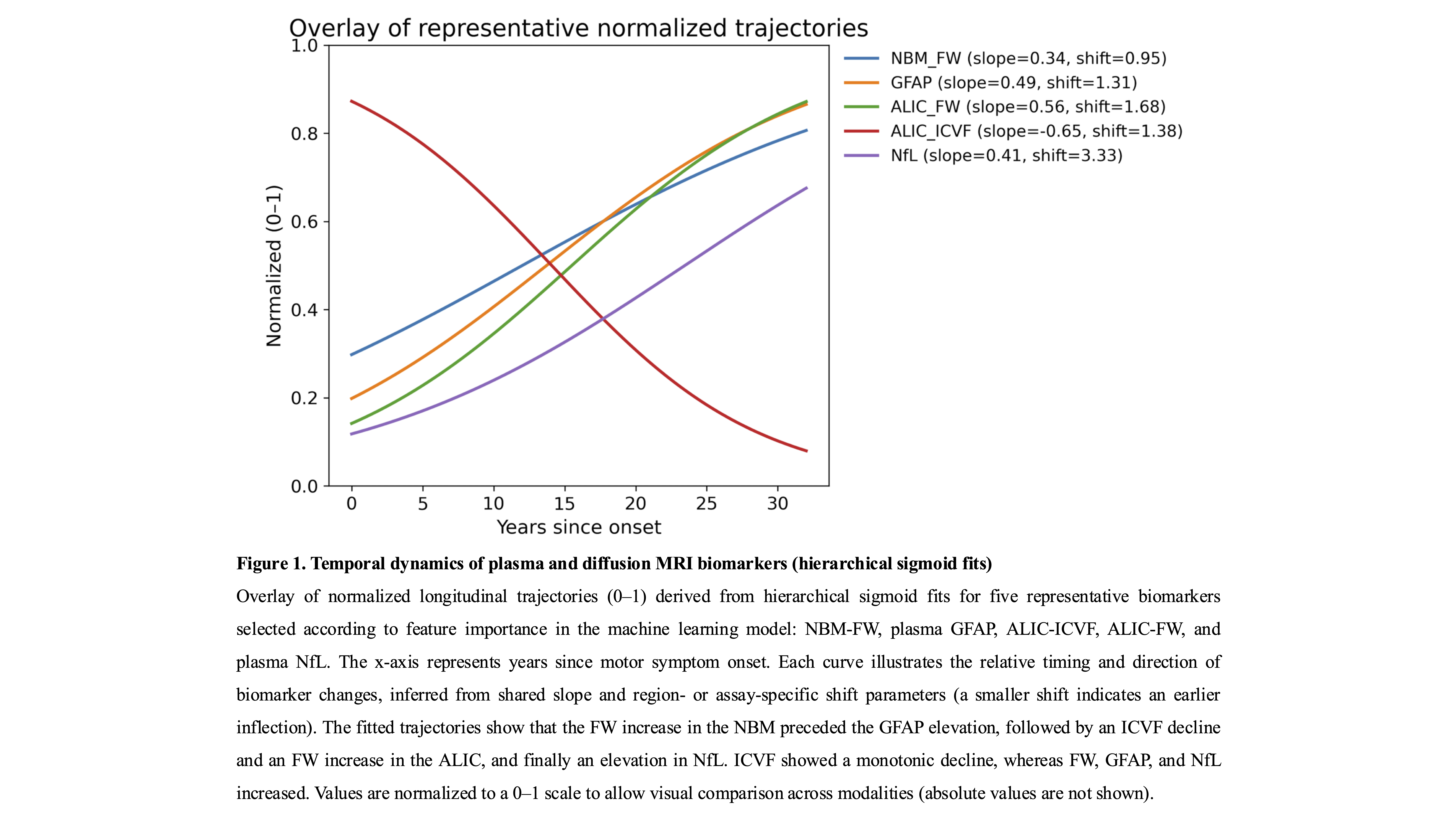
Figure 1
To cite this abstract in AMA style:
S. Ueno, H. Takeshige-Amano, S. Miyaguchi, K. Takabayashi, Y. Yada, Y. Kondo, D. Samayoa, E. Igami, T. Ogawa, S. Ueno, W. Sako, T. Tateishi, T. Hoshino, K. Kamagata, N. Hattori, N. Honda, T. Hatano. Spatiotemporal Trajectories of Plasma Biomarkers and Diffusion MRI Metrics in Parkinson’s Disease [abstract]. Mov Disord. 2026; 41 (suppl 1). https://www.mdsabstracts.org/abstract/spatiotemporal-trajectories-of-plasma-biomarkers-and-diffusion-mri-metrics-in-parkinsons-disease/. Accessed October 1, 2026.« Back to 2026 International Congress
MDS Abstracts - https://www.mdsabstracts.org/abstract/spatiotemporal-trajectories-of-plasma-biomarkers-and-diffusion-mri-metrics-in-parkinsons-disease/