
Category: Ataxia
Objective: To highlight an often overlooked cause of ataxia with hearing impairment.
Background: Superficial siderosis (SS) of the nervous system is caused by chronic hemosiderin deposition due to low-grade bleeding into the subarachnoid space. Classical infratentorial SS (iSS) is most commonly associated with spinal dural pathology where chronic bleeding leads to accumulation of hemosiderin in the posterior fossa and usually along the spine. iSS typically presents as an ataxia disorder with hearing impairment in the fourth to sixth decade of life. Cortical SS (cSS) is a localized form characterized by focal deposits, often asymmetrical, of hemosiderin in the supratentorial cortical sulci. cSS is commonly indicative of an underlying cerebral amyloid angiopathy and presents with spells of focal neurological symptoms and cognitive impairment.
Method: Clinical examination, CSF examination, evoked potentials and a series of structural neuroimaging.
Results: Our patient, 64 year-old male, presented with a year long gradually progressive instability during gait and hearing loss. There was no history of subarachnoidal hemorrhage, head trauma or previous operations. Neurological exam showed cerebellar ataxic gait, dystaxia of the upper and lower limbs and a bilateral intentional tremor. CSF examination revealed xanthochromia and significant elevation of red blood cell count. Auditory evoked potential displayed no response in the central components with eradication of waves IV and V. On T2-weighed and T2-susceptibility-weighted brain MRI, supratentorial and infratentorial hemosiderin deposits were diffusely present and mainly concentrated around the cerebellum, brainstem, and along Sylvian and the interhemispheric fissures. MRI of the whole spine showed the highest concentration hemosiderin deposits around the lower cervical spine. On CT myelogram intrathecal contrast extravasation was observed through a dural defect at T2. Surgical repair is scheduled for the thoracic dural defect at T2.
Conclusion: Our case outlines the importance of considering the diagnosis of SS in patient presenting with ataxia and hearing impairment. Fortunately, with widespread use of MRI, superficial siderosis is increasingly being recognized. The combination of cSS and iSS is, however, still uncommon in literature. Nevertheless, genetic testing of our patient for cerebral amyloid angiopathy is in progress in order to rule out this possible but unlikely combination of etiologies.
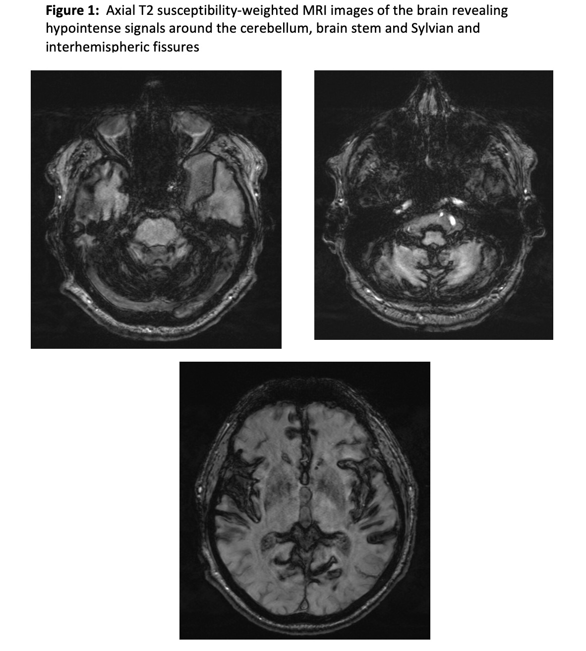
Figue 1
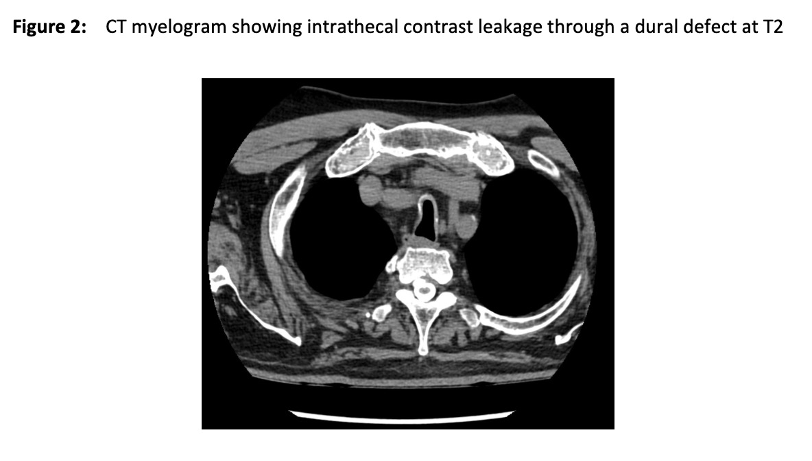
Figure 2
References: Supported by MH CZ – DRO (FNOl, 00098892)
To cite this abstract in AMA style:
ANM. Afifi, L. Satke, E. čecháková. Superficial siderosis; a case review [abstract]. Mov Disord. 2024; 39 (suppl 1). https://www.mdsabstracts.org/abstract/superficial-siderosis-a-case-review/. Accessed November 8, 2026.« Back to 2024 International Congress
MDS Abstracts - https://www.mdsabstracts.org/abstract/superficial-siderosis-a-case-review/