
Category: Parkinson's disease: Neuroimaging
Objective: To compare the performance of 18F-AV133 between NeuroEXPLORER (NX) and standard resolution Siemens Biography (mCT) positron emission tomography (PET) cameras in people with Parkinson’s Disease and Prodromal PD
Background: Loss of dopaminergic neurons in PD begins in the prodromal stage. 18F-AV133, a vesicular monoamine transporter type-2 tracer, is an emerging marker of dopaminergic loss. However, current PET systems have limited resolution and sensitivity, restricting reliable visualization and quantification within the small brainstem nuclei involved in early neurodegeneration, like locus coeruleus (LC) and substantial nigra (SN). NX, a new ultra high resolution PET camera with enhanced sensitivity, may overcome these limitations. Combining the 18F-AV133 with NX has the potential to enhance detection of degenerative changes earlier and more sensitive to detecting decline over time. We present the study design and initial imaging data the ongoing Parkinson’s Progression Markers Initiative (PPMI) substudies.
Method: The PPMI AV133 substudies will enroll prodromal PD, PD, and HC participants. Participants will undergo PET imaging following administration of 6mCi of 18F-AV133. Imaging will be performed using both NX and mCT cameras. Following 10 min acquisition on the first camera [80-90 min post injection (p.i)], the participant will be transferred to 2nd camera for another 10 min acquisition [100 to 110 min p.i]. Imaging will be repeated at year 1 and 2 to compare rate of progression between the two cameras.
Results: Initial enrollment across the two studies include 3 PD, 7 Prodromal PD, and 1 HC. Preliminary imaging results as displayed in Fig 1 and 2 demonstrate improved spatial delineation of small midbrain structures with the NX. LC and SN showed greater contrast and improved visualization with NX compared to mCT allowing quantification otherwise limited. Enrollment and quantitative analyses are ongoing.
Conclusion: The initial imaging data from the ongoing PPMI AV133 substudies reveals that the NX camera enables enhanced visualization and quantification of LC and SN. This advancement could enhance sensitivity in detecting early neurodegenerative and longitudinal changes. Furthermore, the NX will be instrumental in evaluating candidate tracers targeting alpha synuclein and may allow for revisiting tracers targeting pathology or markers localized to smaller nuclei.
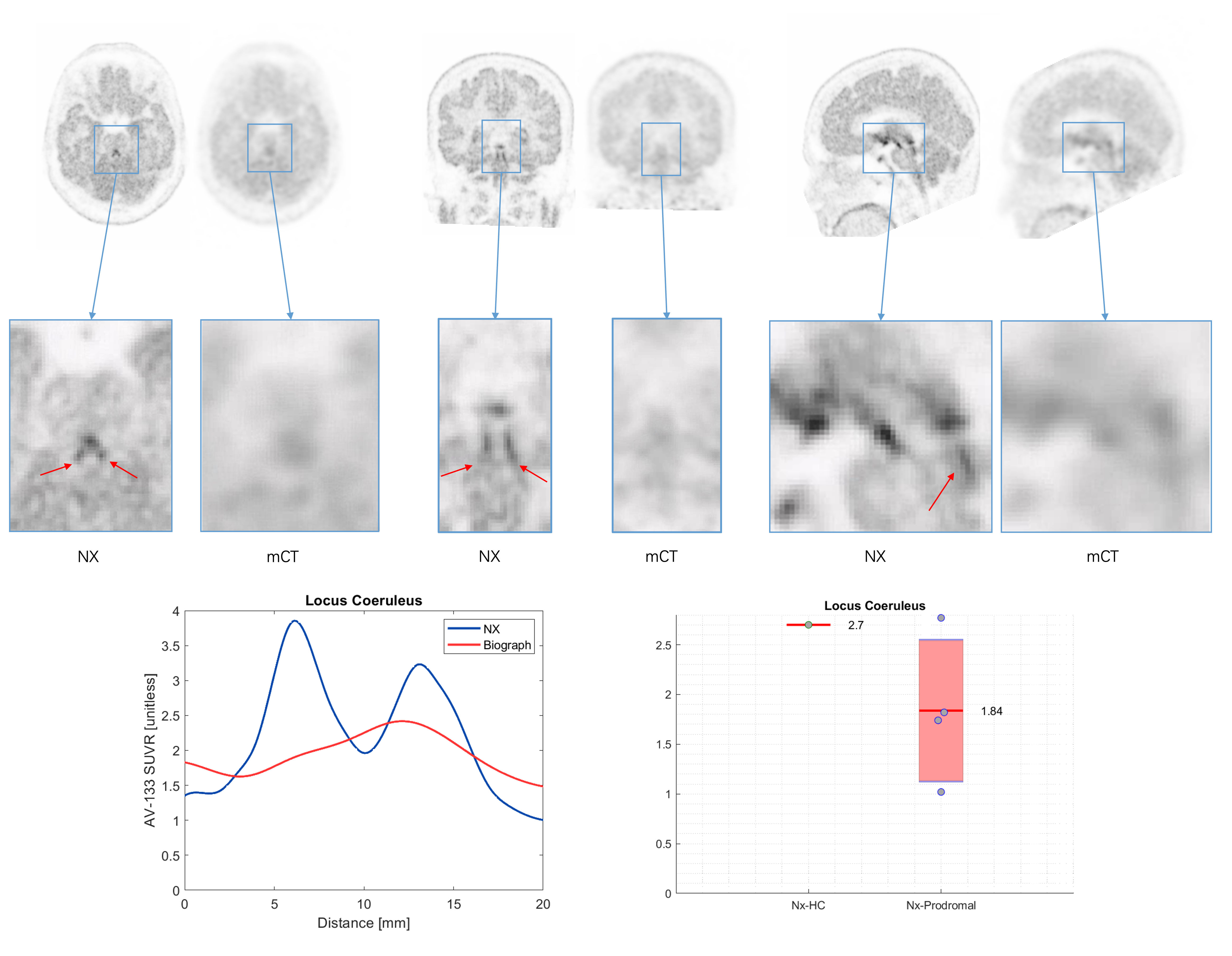
Fig 1: 18F-AV133 imaging on NX and mCT – LC cut.
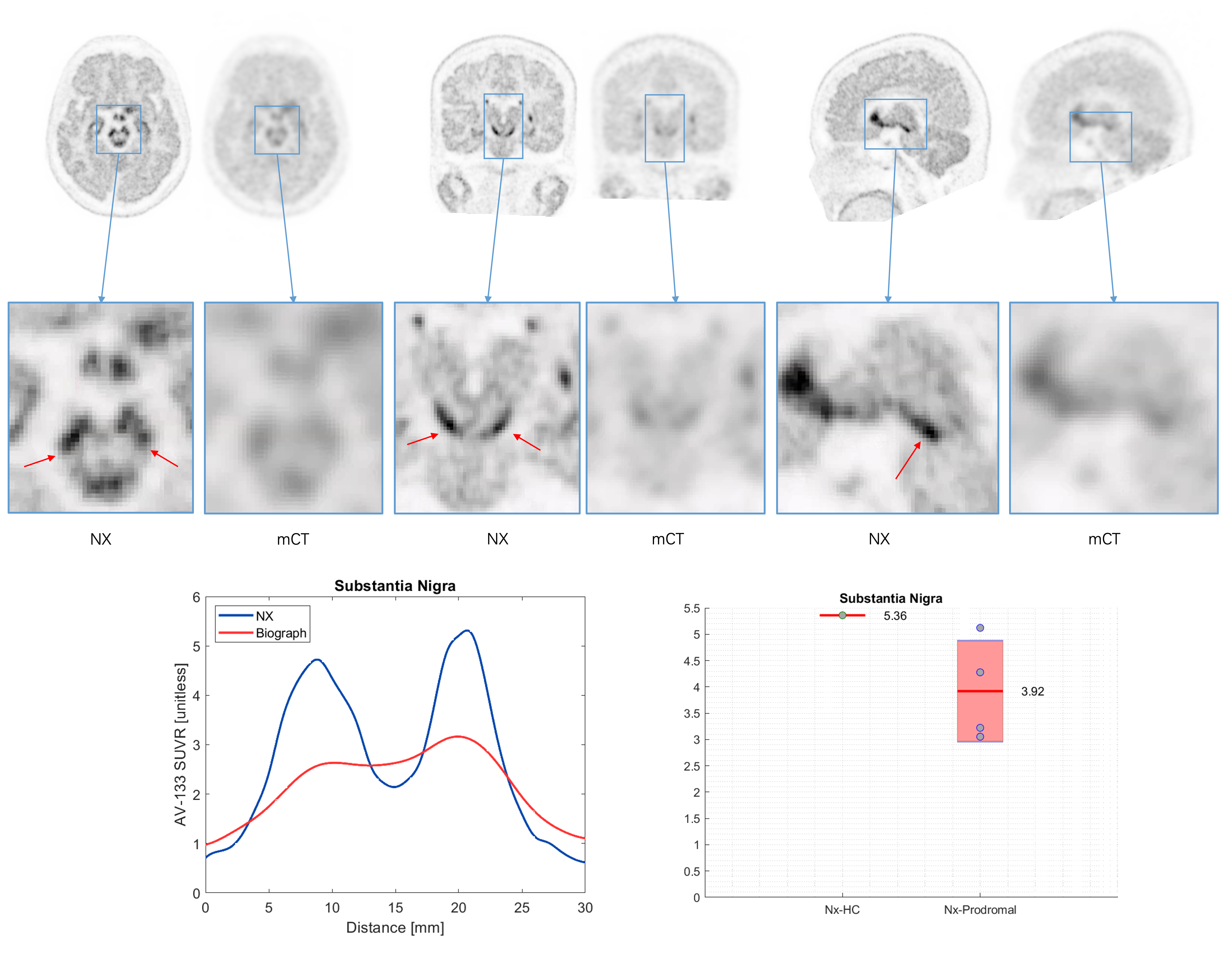
Fig 2: 18F-AV133 imaging on NX and mCT – SN cut.
To cite this abstract in AMA style:
N. Prakash, R. Gunn, G. Rizzo, G. Searle, Z. Fan, J. Eberling, G. Tamagnan, J. Seibyl, K. Marek. Visualization of Small Brainstem Nuclei With the Ultra-High Performance NeuroEXPLORER PET Camera: a Comparison Study Using 18F-AV133 [abstract]. Mov Disord. 2026; 41 (suppl 1). https://www.mdsabstracts.org/abstract/visualization-of-small-brainstem-nuclei-with-the-ultra-high-performance-neuroexplorer-pet-camera-a-comparison-study-using-18f-av133/. Accessed October 1, 2026.« Back to 2026 International Congress
MDS Abstracts - https://www.mdsabstracts.org/abstract/visualization-of-small-brainstem-nuclei-with-the-ultra-high-performance-neuroexplorer-pet-camera-a-comparison-study-using-18f-av133/