
Category: Parkinson's disease: Neuroimaging
Objective: The study aimed to quantify and compare the directional, effective connectivity modulated by HFS and LFS within a motor-cognitive-emotional brain network to identify the frequency-dependent mechanisms underlying improvements in axial symptoms.
Background: While high-frequency stimulation (HFS, >100 Hz) of the subthalamic nucleus (STN) is the standard treatment for Parkinson’s disease (PD), its efficacy for axial symptoms is limited. Low-frequency stimulation (LFS, 60–80 Hz) may offer superior benefits for gait and balance, yet the underlying network-level mechanisms remain poorly understood.
Method: Twenty-one PD patients with bilateral STN-DBS were prospectively enrolled in a randomized, crossover trial. Assessments including MDS-UPDRS-III, Berg Balance Scale, and quantitative gait kinematics were performed under three conditions: HFS (130 Hz), LFS (60 Hz), and off-stimulation. Stochastic dynamic causal modeling (DCM) was applied to resting-state fMRI data to quantify effective connectivity within a network incorporating the basal ganglia (BG), motor thalamus (mThl), mesencephalic locomotor region (MLR), cerebellum (CLBM), motor cortex (M1), and prefrontal regions (DLPFC, ACC).
Results: Both HFS and LFS significantly improved overall motor symptoms (p < 0.001). LFS demonstrated significantly greater improvement than HFS in axial symptoms (p = 0.024), balance (p = 0.014), and gait metrics including speed (p = 0.014) and freezing episodes (p = 0.023). DCM revealed fundamentally distinct modulation: HFS benefits were predominantly mediated by subcortical network enhancement, primarily involving CLBM, mThl, and MLR. Conversely, LFS-induced improvements were specifically correlated with the potentiation of cortico-basal ganglia and cortico-cortical connections, notably between M1, DLPFC, ACC, and BG.
Conclusion: HFS and LFS achieve clinical efficacy via separate network mechanisms: HFS selectively targets subcortical locomotor circuitry, whereas LFS engages cortico-basal ganglia pathways. This dissociation provides a critical foundation for optimizing DBS programming to address refractory axial symptoms in PD.
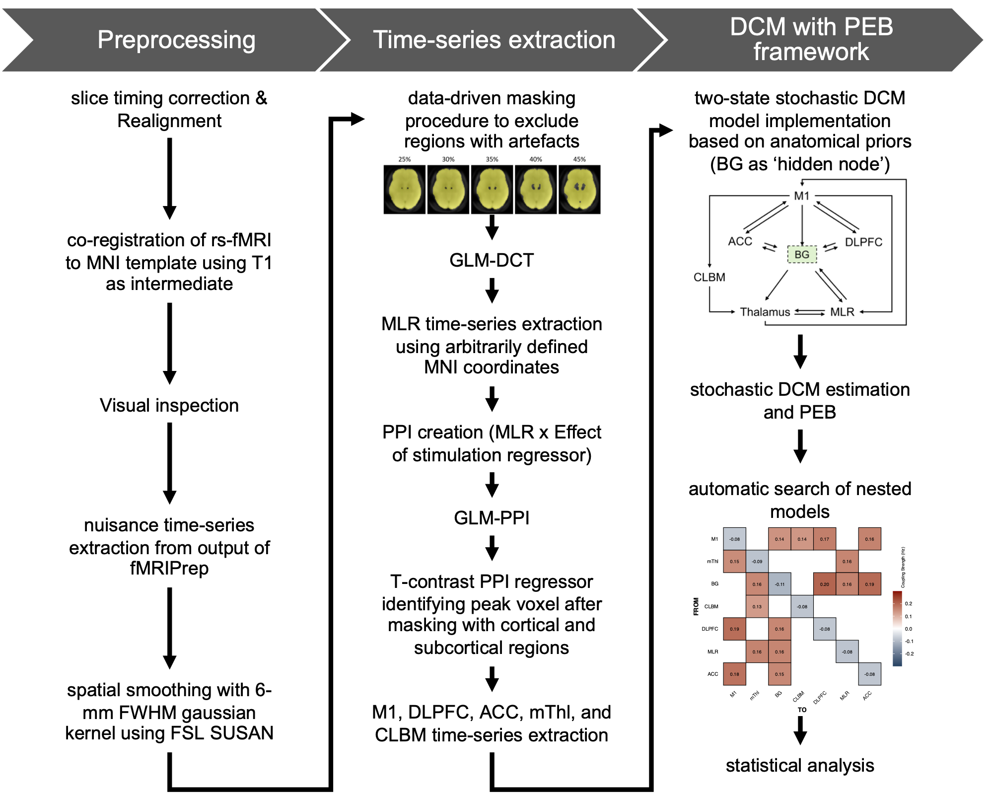
Figure 1.
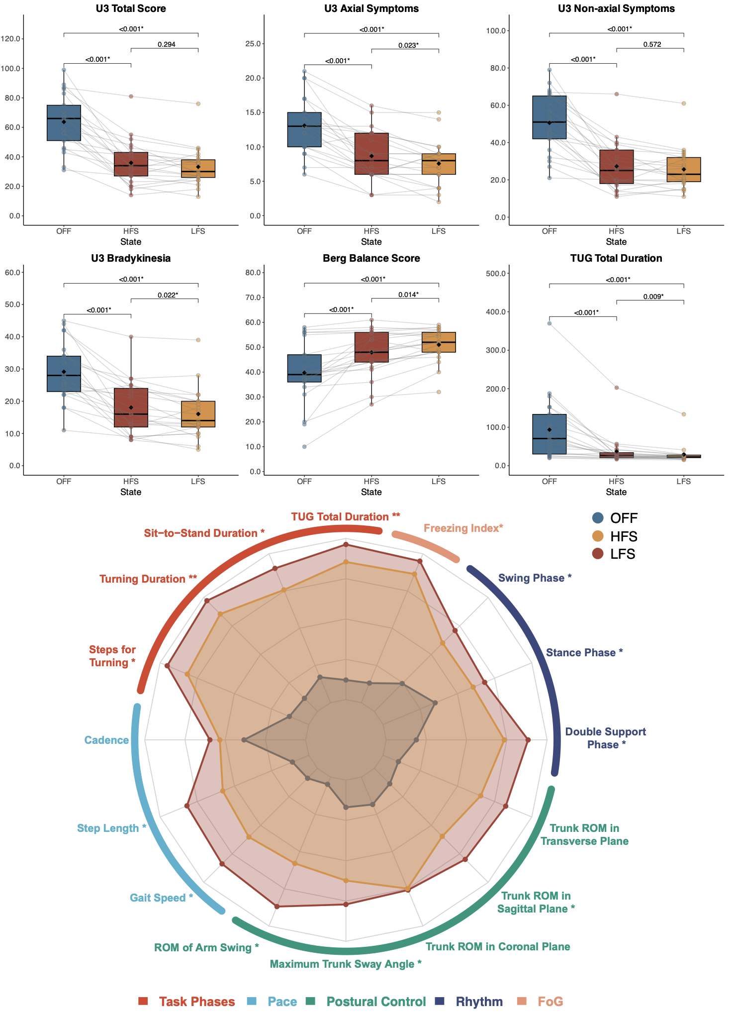
Figure 2.
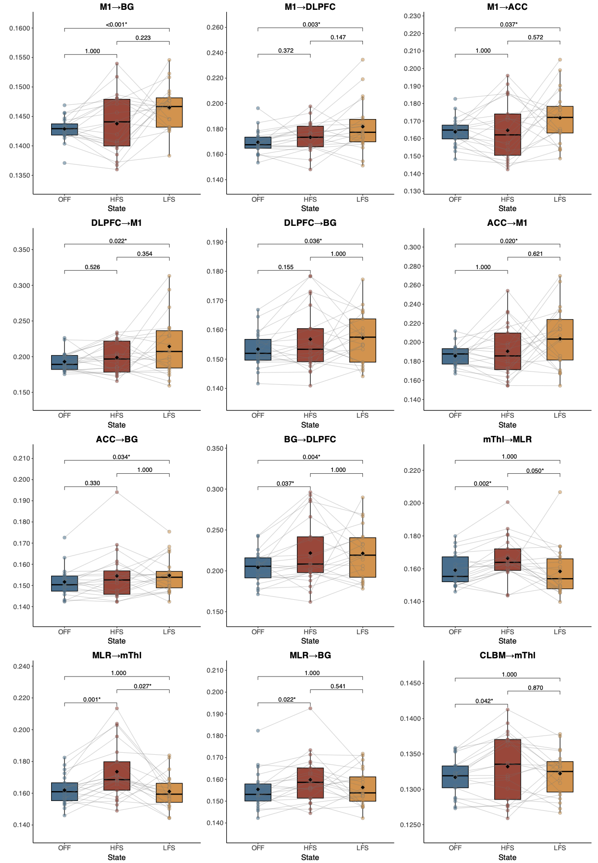
Figure 3.
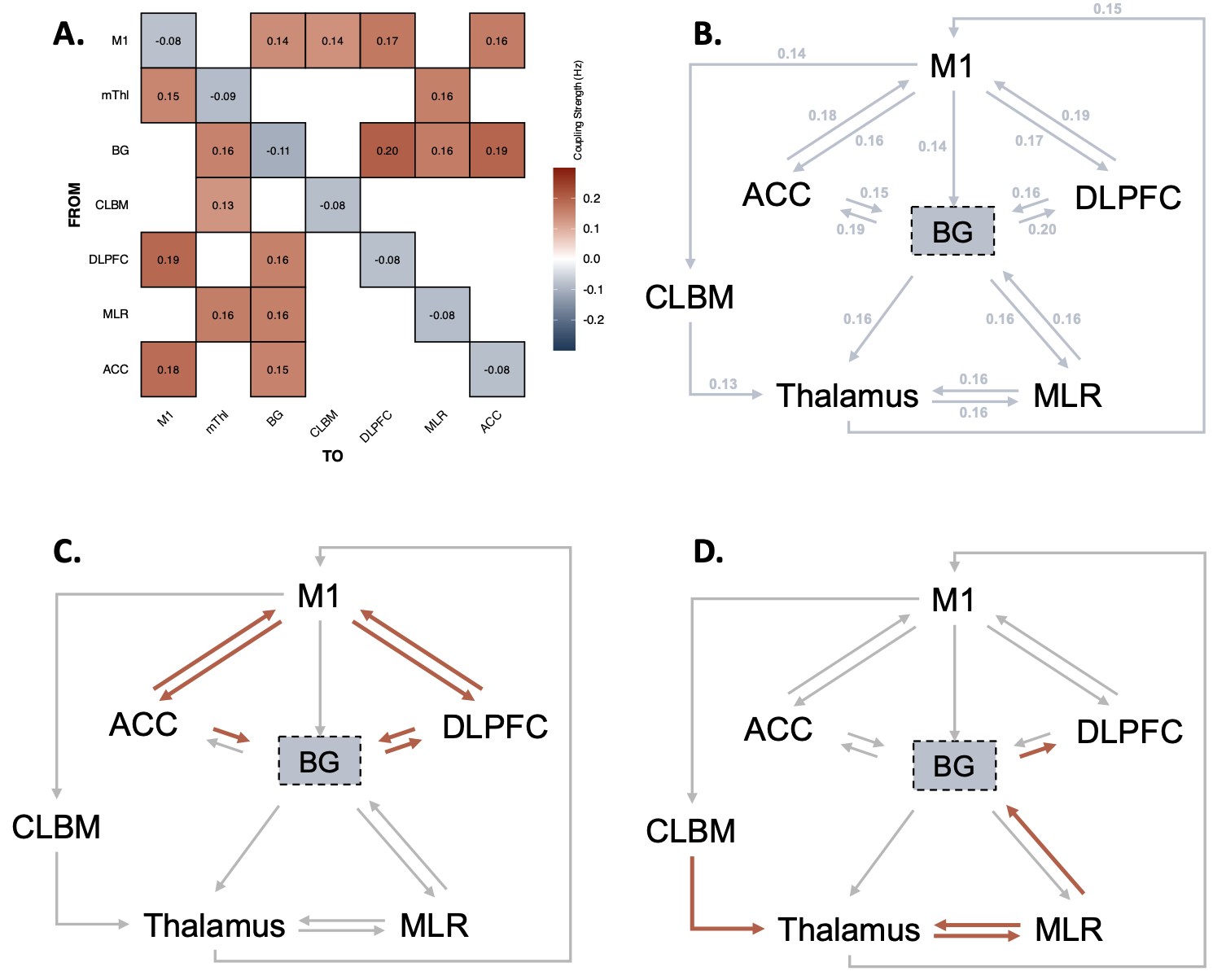
Figure 4.
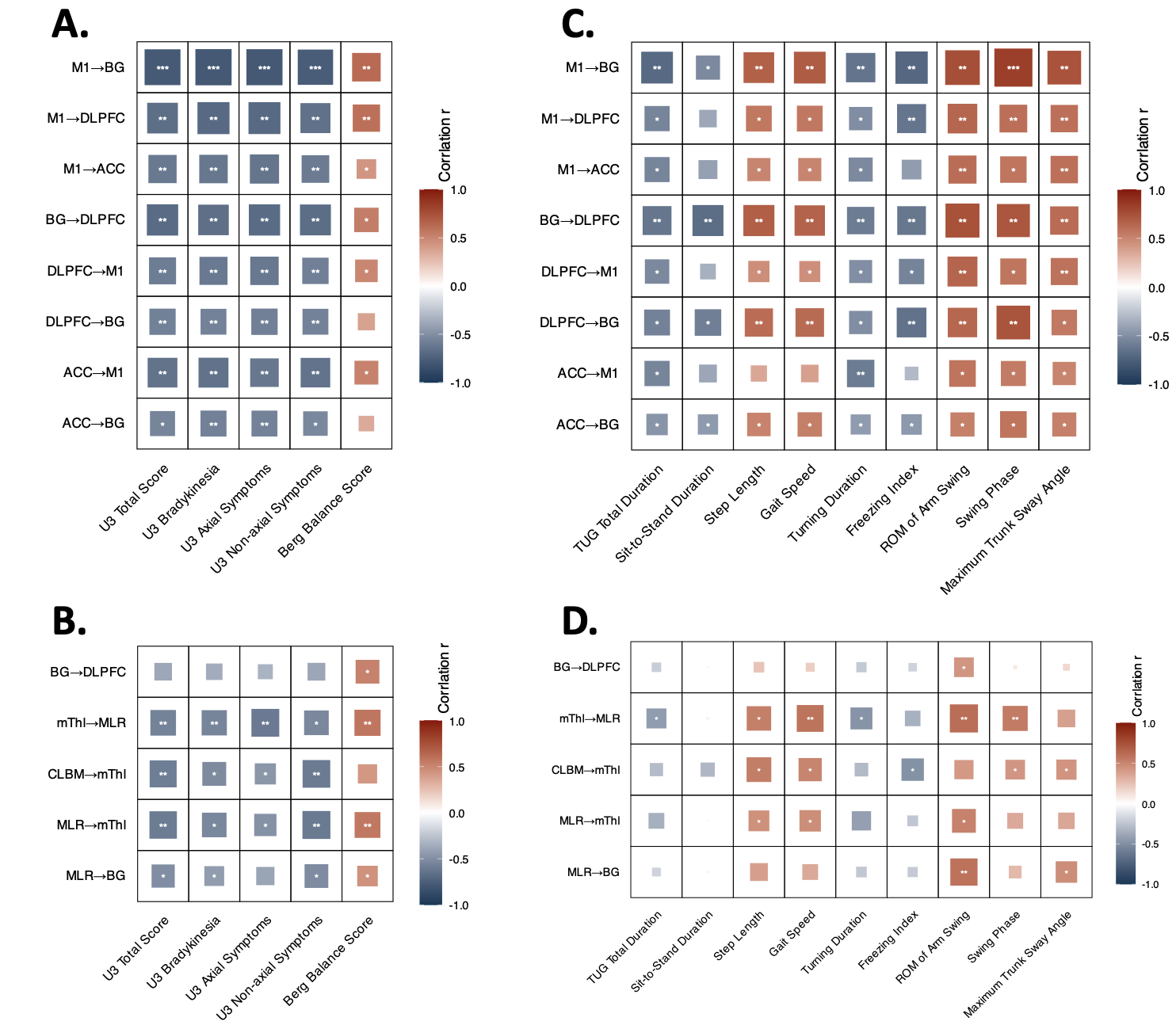
Figure 5.
To cite this abstract in AMA style:
Z. Zeng, Z. Lin, C. Zhang, Z. Duan, B. Sun, N. He, P. Peng, D. Li. Distinct Cortical and Subcortical Network Mechanisms of High and Low Frequency Subthalamic Stimulation in Parkinsons Disease [abstract]. Mov Disord. 2026; 41 (suppl 1). https://www.mdsabstracts.org/abstract/distinct-cortical-and-subcortical-network-mechanisms-of-high-and-low-frequency-subthalamic-stimulation-in-parkinsons-disease/. Accessed October 1, 2026.« Back to 2026 International Congress
MDS Abstracts - https://www.mdsabstracts.org/abstract/distinct-cortical-and-subcortical-network-mechanisms-of-high-and-low-frequency-subthalamic-stimulation-in-parkinsons-disease/