
Category: Parkinson's disease: Neuroimaging
Objective: Diffusion tensor imaging (DTI) holds promising potential for investigating pathological changes in white matter (WM). In recent years, numerous studies worldwide have demonstrated that patients with parkinsonism exhibit microstructural alterations in WM at the early stage. By utilizing DTI to analyze differences in WM microstructure between Parkinson’s disease (PD) and atypical parkinsonism (AP), this study aims to explore the potential value of DTI in the early differential diagnosis of PD and AP.
Background: Parkinsonism is a common clinical manifestation of Parkinson’s disease(PD)and other atypical parkinsonism syndromes(AP), with its core motor symptoms including bradykinesia, resting tremor and rigidity. Diffusion Tensor Imaging(DTI)holds promising application prospects for investigating the pathological changes of cerebral white matter(WM).This study aims to analyze the differences in early white matter microstructural alterations between PD and AP using DTI and to explore the potential value of DTI in the early differential diagnosis of PD and AP.
Method: The researchers collected DTI data from the following study subjects:14 patients with early-stage Parkinson’s disease (PD), 17 patients with middle-to-late-stage PD, 16 patients with atypical parkinsonian syndromes (AP), and 11 age-matched healthy controls (HC). The fractional anisotropy (FA) and apparent diffusion coefficient (ADC) values were measured in the pons, cingulate gyrus, corpus callosum, caudate nucleus, and thalamus.
Results: Statistically significant differences in FA values were observed in the bilateral pons, right caudate nucleus, left thalamus and left substantia nigra among the early PD group, middle-late PD group, AP group and HC group (P<0.05). Meanwhile, Statistically significant differences in ADC values were found in the left pons, corpus callosum, bilateral cingulate gyrus, bilateral caudate nuclei, bilateral thalami, and left substantia nigra (P < 0.05).
Conclusion: Microstructural changes were identified in the pons, corpus callosum, caudate nucleus and thalamus of both PD and AP patients. These changes were more pronounced in early-stage AP than in PD, and were associated with more severe motor symptoms. This difference may be related to neural compensatory mechanisms in early PD involving the cortico–striato–thalamo–cortical (CSTC) circuit and the cerebellar motor circuit.
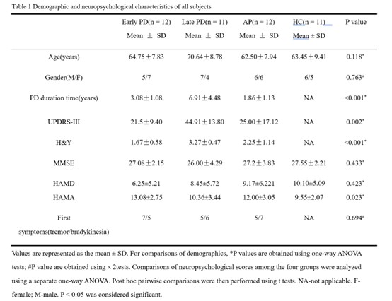
Demographic of all subjects
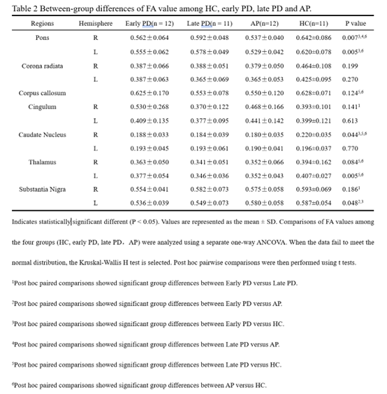
Between-group differences of FA
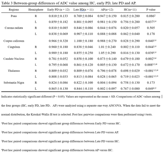
Between-group differences of ADC
To cite this abstract in AMA style:
H. Lin, W. Dai. DTI-Based Comparative Analysis of Microstructural Alterations in Parkinson Disease and Atypical Parkinsonism [abstract]. Mov Disord. 2026; 41 (suppl 1). https://www.mdsabstracts.org/abstract/dti-based-comparative-analysis-of-microstructural-alterations-in-parkinson-disease-and-atypical-parkinsonism/. Accessed October 1, 2026.« Back to 2026 International Congress
MDS Abstracts - https://www.mdsabstracts.org/abstract/dti-based-comparative-analysis-of-microstructural-alterations-in-parkinson-disease-and-atypical-parkinsonism/