
Category: Parkinson's Disease: Surgical Therapy
Objective: To describe DBS electrode localization using Lead-DBS reconstruction and explore its relationship with motor improvement measured by MDS-UPDRS III.
Background: Deep brain stimulation of the subthalamic nucleus (STN-DBS) is an established therapy for Parkinson’s disease (PD). Clinical outcomes depend partly on accurate electrode placement within the dorsolateral motor STN and on optimal postoperative programming. Imaging reconstruction tools such as Lead-DBS allow visualization of DBS electrodes relative to anatomical atlases and may assist clinical decision-making during postoperative programming. Data describing the use of imaging-guided DBS strategies in emerging programs from the Caribbean remain limited.
Method: We performed a prospective single-center case series of PD patients who underwent bilateral STN-DBS implantation. Postoperative imaging was reconstructed using Lead-DBS. Reconstructed DBS leads were classified according to their anatomical location within motor or non-motor STN territories based on atlas segmentation. Patients were categorized as non-motor STN if at least one electrode was located outside the motor STN territory. Motor outcomes were assessed using MDS-UPDRS III before surgery and at 3-month follow-up. Motor improvement was calculated as percentage change from baseline. Changes in levodopa equivalent daily dose (LEDD) were also evaluated. Differences between groups were explored using the Mann–Whitney U test.
Results: Seven patients were analyzed. Mean age at surgery was 62 years and mean disease duration was 9.7 years. Baseline MDS-UPDRS III OFF score was 54.1. At 3 months after STN-DBS, mean motor improvement was approximately 75%. Mean LEDD decreased from 923 mg/day preoperatively to 204 mg/day (≈78% reduction). Three patients had electrodes within the motor STN, while four had at least one electrode in non-motor STN regions. Motor improvement was 78% vs 73.9%, respectively (p=0.63).
Conclusion: Lead-DBS reconstruction confirmed appropriate electrode localization in this emerging DBS program. Substantial motor improvement and marked LEDD reduction were observed, supporting the feasibility of imaging-based DBS verification in the Dominican Republic.
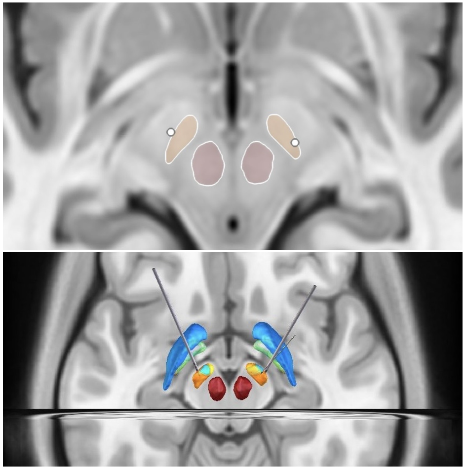
Figure 1. Representative Lead-DBS reconstruction
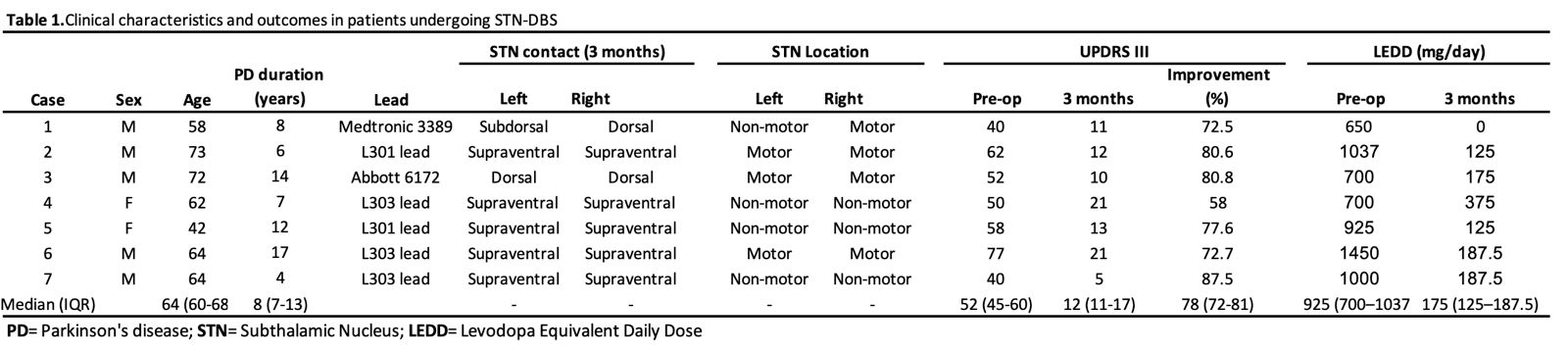
Table 1. Patient characteristics
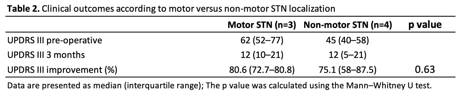
Table 2. Clinical outcome
References: 1. Waldthaler, J. et al. Imaging-based programming of subthalamic nucleus deep brain stimulation in Parkinson’s disease. Brain Stimul. 14, 1109–17; 10.1016/j.brs.2021.07.064 (2021).
2. Pavese, N., Tai, Y. F., Yousif, N., Nandi, D. & Bain P. G. Traditional Trial and Error versus Neuroanatomic 3-Dimensional Image Software-Assisted Deep Brain Stimulation Programming in Patients with Parkinson Disease. World Neurosurg. 134, e98–102; 10.1016/j.wneu.2019.09.106 (2020).
3. Torres V, Del Giudice K, Roldán P, Rumià J, Muñoz E, Cámara A, Compta Y, Sánchez-Gómez A, Valldeoriola F. Image-guided programming deep brain stimulation improves clinical outcomes in patients with Parkinson’s disease. NPJ Parkinsons Dis. 2024 Jan 27;10(1):29. doi: 10.1038/s41531-024-00639-9. PMID: 38280901; PMCID: PMC10821897.
To cite this abstract in AMA style:
L. Reyes Lagarde, C. Ramirez Mateo, I. Suero Matos, V. Torres, A. Perez Fernandez, A. Carreras, K. Del Giudice. Electrode localization in STN deep brain stimulation using image reconstruction from an emerging DBS program in the Dominican Republic [abstract]. Mov Disord. 2026; 41 (suppl 1). https://www.mdsabstracts.org/abstract/electrode-localization-in-stn-deep-brain-stimulation-using-image-reconstruction-from-an-emerging-dbs-program-in-the-dominican-republic/. Accessed October 1, 2026.« Back to 2026 International Congress
MDS Abstracts - https://www.mdsabstracts.org/abstract/electrode-localization-in-stn-deep-brain-stimulation-using-image-reconstruction-from-an-emerging-dbs-program-in-the-dominican-republic/