
Category: Parkinson's disease: Neuroimaging
Objective: Determine whether baseline Diffusion Tensor Imaging (DTI) measures of the substantia nigra (SN) predict fall incidence and burden in Parkinson’s disease (PD).
Background: Degeneration of dopaminergic neurons in the SN in PD is associated with bradykinesia, rigidity, resting tremor, and gait impairment [1,2], which may contribute to falls; affecting the quality of life and increasing the risk of hospitalization and morbidity [3,4]. Falls have been associated with pathophysiological changes observed on imaging [5]. DTI has been used to map and quantify microstructural integrity of white matter tracts in the SN and corpus callosum (CC) of PD patients [6,7,8]. DTI measures in the CC and Pedunculopontine Nucleus are associated with fall burden [9], postural instability, and gait difficulty, key contributors to falls [10]. We hypothesized that DTI measures of the SN could predict future fall incidence and burden in Parkinson’s Progression Markers Initiative (PPMI) participants with PD.
Method: Standardized fractional anisotropy (FA), mean (MD), radial (RD), and axial (AD) diffusivity were calculated for the SN regions of interest [11,12] and global total average for 106 PPMI [11] participants with baseline DTI. Data analysis was restricted to those without falls at baseline by removing those with positive “fall outcomes” at baseline. “Fall outcomes” were also calculated for years 6-14 after baseline from “Determination of Freezing and Falls” items 2, 4-6 with scores ≥1 equaling a fall. Fall incidence (FI) and Fall Burden (FB) were defined as having at least one reported fall and the average calculated score, respectively. We performed linear regressions for FI and FB and ROC analysis for FI with DTI, sex, and age at enrollment as predictors. Successful predictive performance was defined as ROC >90%.
Results: There was no difference in median diffusivity values of the SN between participants with or without falls. Linear regression models did not show significant associations in these measures (Table 1). ROC models showed no discrimination for FI (Table 2).
Conclusion: Contrary to our hypothesis, baseline SN DTI measures were not significantly different between those who fell and those who did not, nor were they predictive of future FI or FB. This highlights the importance of seeking alternative predictive biomarkers if further studies confirm this lack of association.
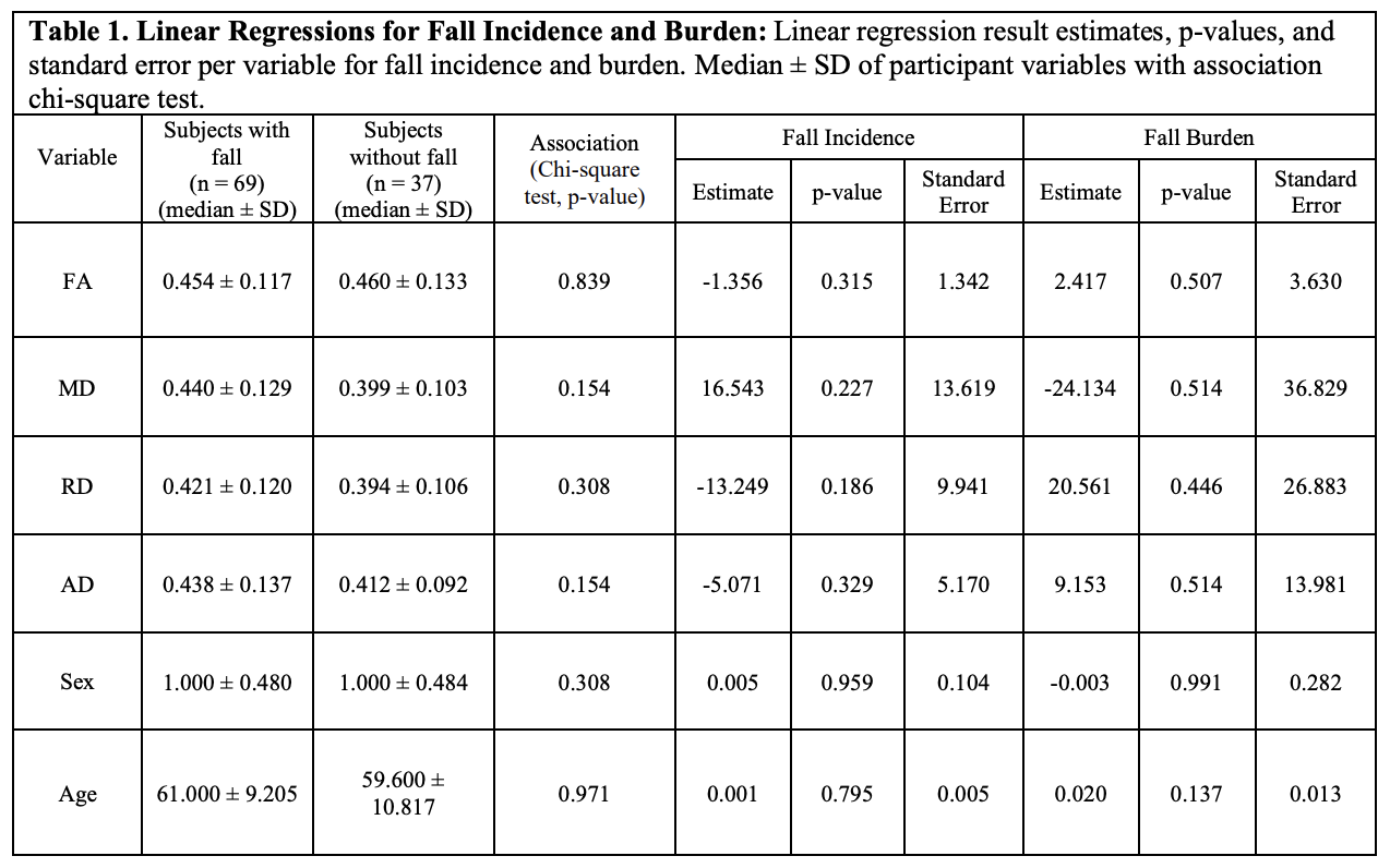
Linear Regressions for Fall Incidence and Burden
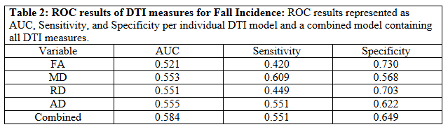
ROC results of DTI measures for Fall Incidence
References: 1. Kalia LV, Lang AE. Parkinson’s disease. Lancet. 2015;386(9996):896-912. doi:10.1016/S0140-6736(14)61393-3
2. Mirelman A, Bonato P, Camicioli R, et al. Gait impairments in Parkinson’s disease. Lancet Neurol. 2019;18(7):697-708. doi:10.1016/S1474-4422(19)30044-4 3. Murueta-Goyena, A., Muiño, O. & Gómez-Esteban, J.C. Prognostic factors for falls in Parkinson’s disease: a systematic review. Acta Neurol Belg 124, 395–406 (2024). https://doi.org/10.1007/s13760-023-02428-2
4. Zhang Y, Zhang Y, Yan Y, Kong X, Su S. Risk factors for falls in Parkinson’s disease: a cross-sectional observational and Mendelian randomization study. Front Aging Neurosci. 2024;16:1420885. doi:10.3389/fnagi.2024.1420885
5. Bae, Yun Jung et al. “Imaging the Substantia Nigra in Parkinson Disease and Other Parkinsonian Syndromes.” Radiology vol. 300,2 (2021): 260-278. doi:10.1148/radiol.2021203341
6. Zhang, Yu, and Marc A. Burock. “Corrigendum: Diffusion Tensor Imaging in Parkinson’s Disease and Parkinsonian Syndrome: A Systematic Review.” Frontiers in Neurology, vol. 11, 2020, Frontiers Media SA, https://doi.org/10.3389/fneur.2020.612069
7. Yang, K., Wu, Z., Long, J. et al. White matter changes in Parkinson’s disease. npj Parkinsons Dis. 9, 150 (2023). https://doi.org/10.1038/s41531-023-00592-z
8. Chan LL, Ng KM, Rumpel H, Fook-Chong S, Li HH, Tan EK. Transcallosal diffusion tensor abnormalities in predominant gait disorder parkinsonism. Parkinsonism Relat Disord. 2014;20(1):53-59. doi:10.1016/j.parkreldis.2013.09.017
9. Craig CE, Jenkinson NJ, Brittain JS, et al. Pedunculopontine Nucleus Microstructure Predicts Postural and Gait Symptoms in Parkinson’s Disease. Mov Disord. 2020;35(7):1199-1207. doi:10.1002/mds.28051
10. Liu WY, Tung TH, Zhang C, Shi L. Systematic review for the prevention and management of falls and fear of falling in patients with Parkinson’s disease. Brain Behav. 2022;12(8):e2690. doi:10.1002/brb3.2690.
11. Parkinson Progression Marker Initiative. The Parkinson Progression Marker Initiative (PPMI). Prog Neurobiol. 2011;95(4):629-635. doi:10.1016/j.pneurobio.2011.09.005
12. O’Donnell, L. J., & Westin, C. F. (2011). An introduction to diffusion tensor image analysis. Neurosurgery clinics of North America, 22(2), 185–viii. https://doi.org/10.1016/j.nec.2010.12.004
To cite this abstract in AMA style:
I. Bedoy, J. Tejeda, R. Rajmohan, N. Phielipp. Investigating Substantia Nigra Microstructural Integrity as a Potential Predictive Biomarker for Fall Burden in Parkinson’s Disease [abstract]. Mov Disord. 2026; 41 (suppl 1). https://www.mdsabstracts.org/abstract/investigating-substantia-nigra-microstructural-integrity-as-a-potential-predictive-biomarker-for-fall-burden-in-parkinsons-disease/. Accessed October 1, 2026.« Back to 2026 International Congress
MDS Abstracts - https://www.mdsabstracts.org/abstract/investigating-substantia-nigra-microstructural-integrity-as-a-potential-predictive-biomarker-for-fall-burden-in-parkinsons-disease/