
Category: Parkinson's disease: Neuroimaging
Objective: This study characterizes the microstructural evolution of the locus coeruleus (LC) and its thalamic projection tract in Parkinson’s disease (PD) using free‑water imaging across two cohorts. We evaluated longitudinal changes in LC integrity, examined the coupling between LC degeneration and its projection fibers, and assessed tract‑related effects on 14 thalamic subregions. Mediation analysis was applied to test whether LC–thalamic fibers mediate the impact of LC damage on the centromedian–parafascicular complex.
Background: The locus coeruleus (LC) projects widely throughout the brain, with the thalamus receiving its densest noradrenergic input. The LC–thalamic tract serves as a central pathway linking the LC to the cortex and is a key component of the noradrenergic system1. Using 7T MRI, Wanders et al. reported early Parkinson’s disease (PD) patients show widespread white matter decline and specifically examined LC–thalamic fibers, highlighting their association with early non‑motor symptoms of PD2.
Method: Cohort 1 included PD patients recruited at Guangdong Provincial People’s Hospital (2021–2025) with clinical and MRI data acquired on a Philips Ingenia 3.0T system. Cohort 2 data were obtained from the PPMI database (Siemens Trio‑tim 3T) under authorization. LC microstructural analysis was performed using the 7T MRI probabilistic mapping method of Ye et al3.
Results: Cohort 1 (263 PD, 46 HC) and Cohort 2 (126 PD, 49 HC) were well matched. LC integrity declined across disease stages in Cohort 1, while longitudinal analysis in Cohort 2 showed increasing LC MDt (β = 0.102, P = 0.044) over 4 years. LC damage correlated with LC–thalamic tract degeneration (P < 0.05), and tract integrity measures (FWf, FAt, MD) were associated with microstructural changes in most thalamic subregions, especially the CM‑Pf (P < 0.001). Mediation analysis (FDR‑corrected) confirmed that the LC–thalamic tract significantly mediates the impact of LC degeneration on the thalamic CM‑Pf complex.
Conclusion: In conclusion, the LC–thalamic circuit shows progressive microstructural degeneration in PD, with longitudinal LC damage strongly coupled to its projection fibers. Mediation analysis demonstrates that the LC–thalamic tract transmits pathological effects from the brainstem to thalamic subregions, particularly the CM‑Pf, underscoring anatomical connectivity as a key pathway in PD progression.
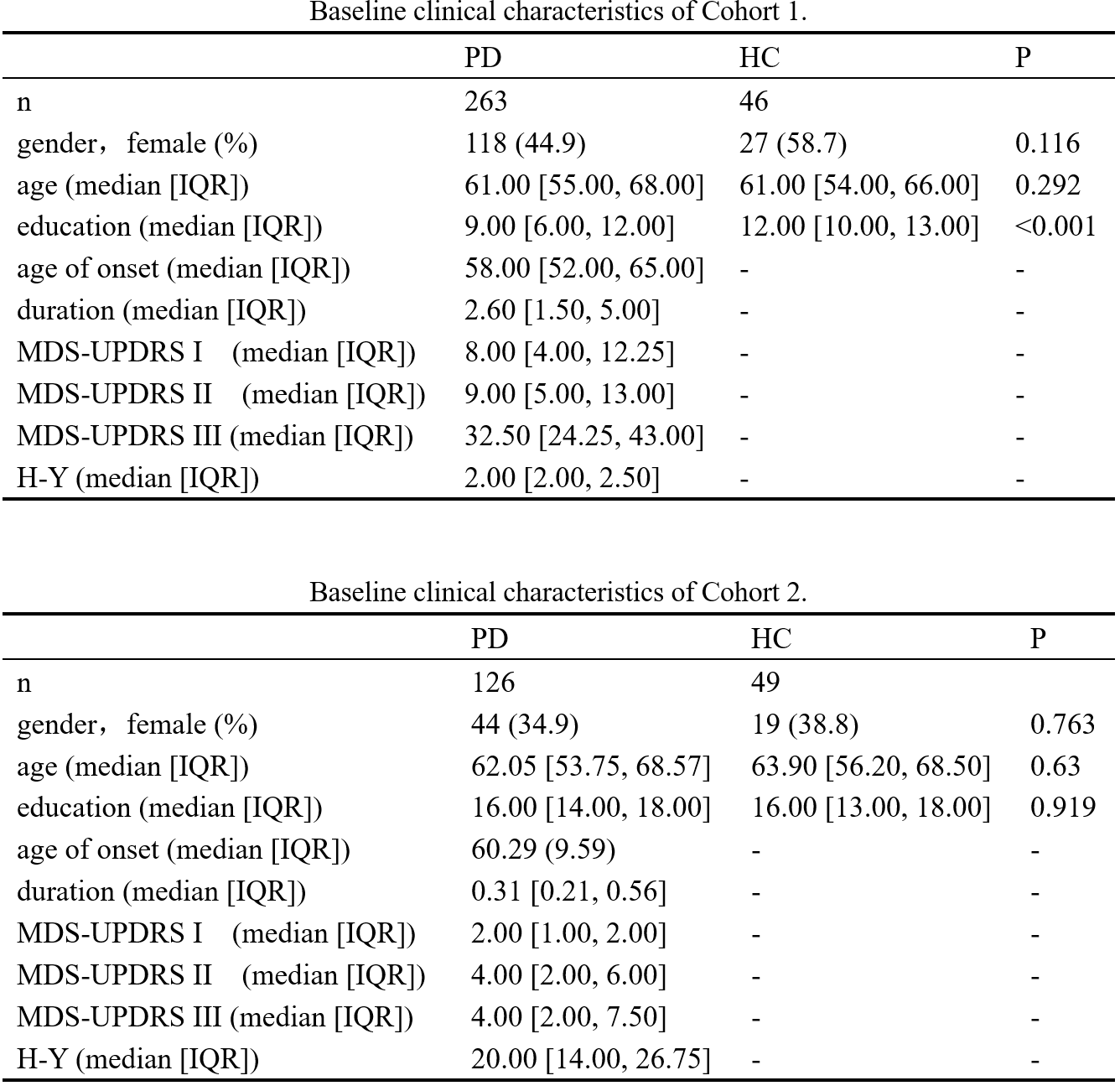
Table1.
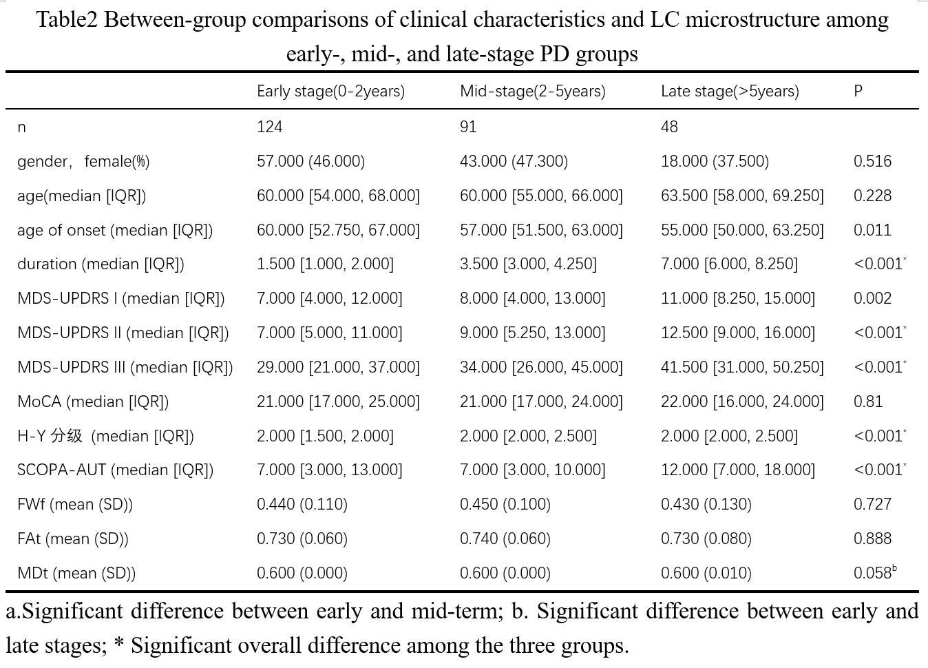
Table2.
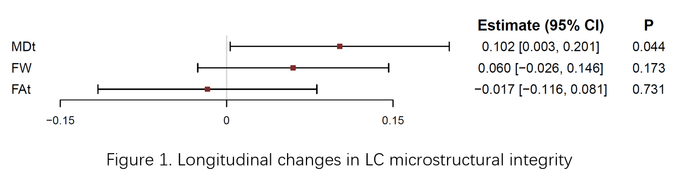
Figure 1.
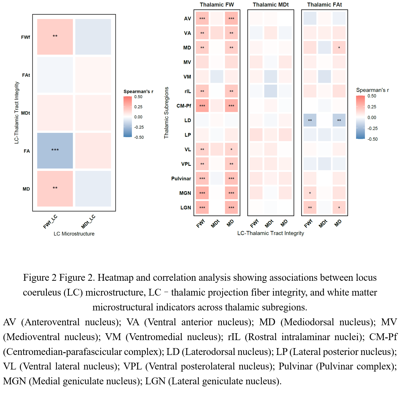
Figure 2.
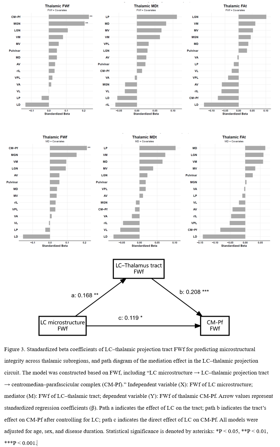
Figure3.
References: 1. Hwang, K., Bertolero, M. A., Liu, W. B. & D’Esposito, M. The Human Thalamus Is an Integrative Hub for Functional Brain Networks. J. Neurosci. 37, 5594–5607 (2017).
2. Wanders, M. G. et al. Altered white matter connection from the locus coeruleus to the thalamus in Parkinson’s disease. Neuroimage Rep. 5, 100292 (2025).
3. Ye, R. et al. An in vivo probabilistic atlas of the human locus coeruleus at ultra-high field. NeuroImage 225, 117487 (2021).
To cite this abstract in AMA style:
XL. Cui, MF. Cai, YH. Zhang. Locus Coeruleus-Thalamic Circuit Degeneration in Parkinson’s Disease [abstract]. Mov Disord. 2026; 41 (suppl 1). https://www.mdsabstracts.org/abstract/locus-coeruleus-thalamic-circuit-degeneration-in-parkinsons-disease/. Accessed October 1, 2026.« Back to 2026 International Congress
MDS Abstracts - https://www.mdsabstracts.org/abstract/locus-coeruleus-thalamic-circuit-degeneration-in-parkinsons-disease/