
Objective: To illustrate the utility of EMG/NCS in diagnosing a paroxysmal craniocervical dystonia confounded by features mimicking seizure and functional movement disorder (FMD).
Background: A 66-year-old woman presented with three years of episodic facial movements. Symptoms involved muscle contraction sequentially involving left eye closure, right eye closure, partial mouth closure with tongue protrusion, and activation of neck muscles. Craniocervical dystonia was initially considered, but various features drew pause: paroxysmal events with a late age of onset, eye closure that did not appear forcibly closed, painlessness, lack of response to a sensory trick, and no significant response to Botox. She described preceding abdominal discomfort and left facial numbness, suggesting seizure, but video-EEG capturing the episodes lacked ictal correlate. FMD was considered due to fluctuations (opening eyes mid-event) and suggestibility (delaying onset until witnessed).
Method: Multi-channel EMG recordings were performed of the left and right orbicularis oculi, left SCM, left submental region, left lower abdomen, and left extensor indicis. Recordings took place at rest, with multiple volitional movements, and during three symptomatic episodes.
Results: Muscle firing began in the left orbicularis oculi with rapid spread to the right orbicularis oculi. Firing was tonic, continuous, lasting for several seconds, and followed by caudal spread to the SCM/hypoglossal region. After a gradual decrease of firing, an intermittent rhythmic resting tremor of 5-6 Hz in the left extensor indicis emerged. Bilaterally, there was a lack of blink reflex habituation up to a frequency of 3Hz. Volitional left hand grip triggered low-grade firing in the left facial muscles. Volitional movements of the right fingers triggered brief motor unit action potentials in the resting left extensor indicis at a frequency of 2-3Hz.
Conclusion: The ocular features were clarified as blepharospasm supported by impaired blink reflex habituation [1-2]. Mirroring and motor overflow were identified, and at times were without a clinical correlate [3]. The case highlights the utility of EMG/NCS to dysambiguate clinical findings and reveal subclinical neuromuscular electrographic features to solidify a diagnosis of dystonia.
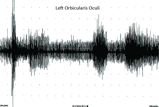
Dystonic firing pattern
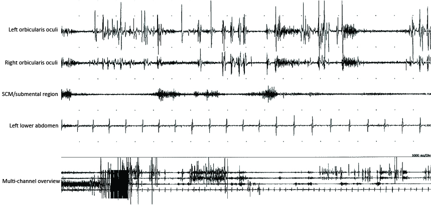
Rostrocaudal evolution of muscle firing
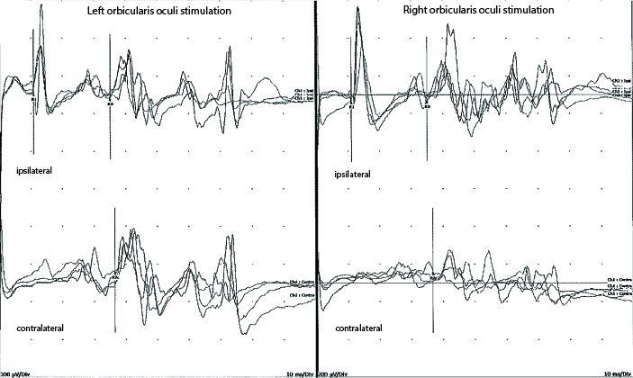
Impaired blink reflex habituation
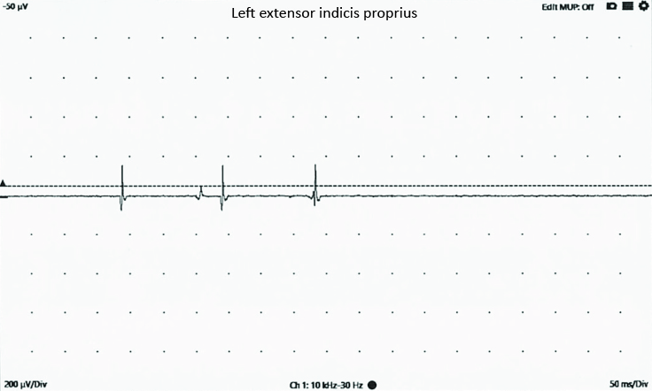
Mirror phenomenon
References: [1] Kimura J, Lyon LW. Orbicularis oculi reflex in the Wallenberg syndrome: alteration of the late reflex by lesions of the spinal tract and nucleus of the trigeminal nerve. Journal of Neurology, Neurosurgery & Psychiatry. 1972;35(2):228–233.
[2] Berardelli A, Rothwell JC, Day BL, Marsden CD. Pathophysiology of blepharospasm and oromandibular dystonia. Brain. 1985;108(3):593–608.
[3] Quattrone A, Latorre A, Magrinelli F, Mulroy E, Rajan R, Neo RJ, Rothwell JC, Bhatia KP. A reflection on motor overflow, mirror phenomena, synkinesia and entrainment. Movement Disorders Clinical Practice. 2023;10(9):1243–1252.
To cite this abstract in AMA style:
A. Talaugon*, A. Young*, S. Jaradeh, N. Samudra, B. Razavi, P. Kahali. The Role of Electrophysiology in Preventing Diagnostic Misclassification of a Craniocervical Dystonia [abstract]. Mov Disord. 2026; 41 (suppl 1). https://www.mdsabstracts.org/abstract/the-role-of-electrophysiology-in-preventing-diagnostic-misclassification-of-a-craniocervical-dystonia/. Accessed October 1, 2026.« Back to 2026 International Congress
MDS Abstracts - https://www.mdsabstracts.org/abstract/the-role-of-electrophysiology-in-preventing-diagnostic-misclassification-of-a-craniocervical-dystonia/