Session Information
Date: Monday, October 8, 2018
Session Title: Parkinson's Disease: Neuroimaging And Neurophysiology
Session Time: 1:15pm-2:45pm
Location: Hall 3FG
Objective: To identify neural correlates of mild cognitive impairment in Parkinson’s disease (PD-MCI) according to Level-I Movement Disorders Task Force Criteria (MDS-TF) (1), using a multimodal approach that combines structural, functional and topological measures of neurocognitive networks by Voxel-Based Morphometry (VBM), Functional Connectivity (FC) and Graph Theory (GT).
Background: Neurocognitive networks (Salience, Default, Central Executive) have shown early connectivity changes in several neurodegenerative diseases (2). FC and GT could help to identify early disruption in large-scale networks of PD patients with normal cognition (PD-NC) and PD-MCI before structural damage occurs.
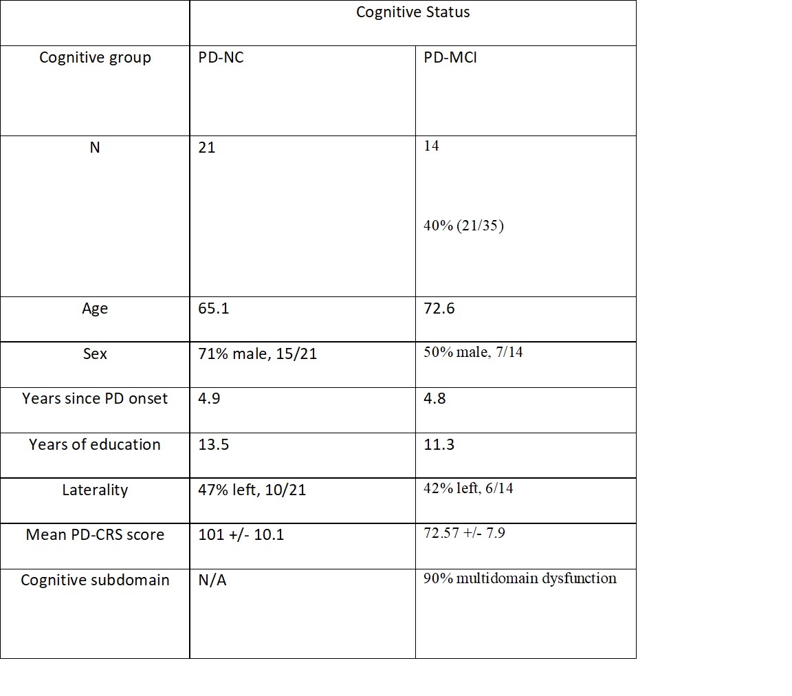
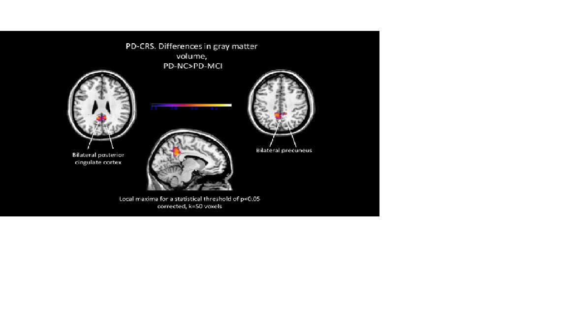
Methods: Prospective study of 35 consecutive non-demented PD patients classified for cognitive impairment using Level-I MDS-TF criteria. Cognitive subdomains were assessed using Level-II criteria. VBM, FC and GT measures were obtained from a 3T-MRI scanner, pre-processed and analyzed using SPM/CONN toolboxes running in MATLAB. For VBM, a threshold of k=50 contiguous voxels was considered significant. A statistical threshold of p<0.05, FDR-corrected was employed in all analysis.
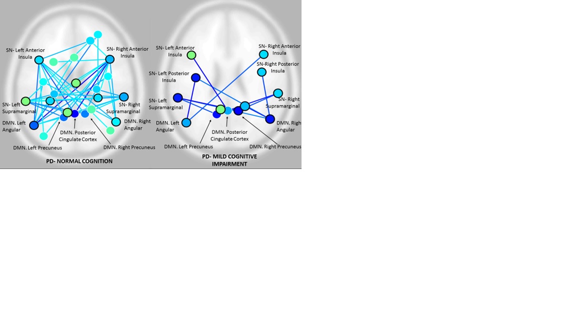
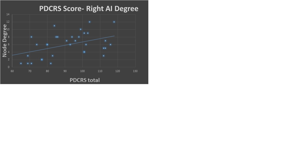
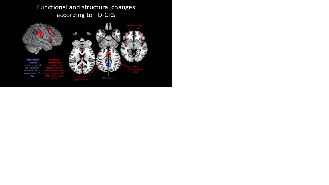
Results: Twenty-one patients were classified as PD-NC, and 14 as PD-MCI. Loss of GMV in bilateral precuneus (DMN) was the main structural difference between groups, while FC and GT differences were restricted to the right Anterior Insula (AI) and right Supramarginal Gyrus (SmG), hubs of the Salience Network. Node degree in right AI significantly correlated with PD-CRS total score and performance in executive tasks, and node degree in right SmG correlated with visuospatial scores.
Conclusions: 1. PD-MCI is associated with both functional and structural changes in neurocognitive networks, showing loss of FC and topological features in Salience Network regions in absence of structural damage, and early GMV loss in bilateral precuneus. 2. These results suggest a loss of compensation mechanisms in Salience Network hubs as one of the first network failures in PD-MCI.
References: 1. Litvan I, et al. Diagnostic Criteria for Mild Cognitive Impairment in Parkinson´s Disease: Movement Disorders Task Force Guidelines. Mov.Disord.2012;27: 349-356. 2. Seeley W, et al. Neurodegenerative diseases target large-scale human brain networks. Neuron 2009; 62:42-52.
To cite this abstract in AMA style:
I. Aracil, F. Sampedro, J. Marin, J. Perez, H. Bejr-Kasem, S. Martinez-Horta, A. Horta, M. Boti, J. Pagonabarraga, J. Kulisevsky. Cognitive networks in Parkinson’s Disease Mild Cognitive Impairment: Salience Network disruption without gray matter loss [abstract]. Mov Disord. 2018; 33 (suppl 2). https://www.mdsabstracts.org/abstract/cognitive-networks-in-parkinsons-disease-mild-cognitive-impairment-salience-network-disruption-without-gray-matter-loss/. Accessed December 24, 2025.« Back to 2018 International Congress
MDS Abstracts - https://www.mdsabstracts.org/abstract/cognitive-networks-in-parkinsons-disease-mild-cognitive-impairment-salience-network-disruption-without-gray-matter-loss/

