Category: MSA, PSP, CBS: Neuroimaging
Objective: To identify the regions with sensitive tau deposition for differentiating progressive supranuclear palsy (PSP) from healthy controls. Additionally, to explore the combination of 18F-APN-1607 for detecting tau aggregates and 18F-FP-DTBZ positron emission tomography (PET) for assessing dopaminergic disruption.
Background: As a neurodegenerative disorder characterized by pathological tau protein deposition, tau deposition in PSP can be evaluated by tau PET. Meanwhile, nigrostriatal presynaptic imaging is used to assess dopaminergic impairment in PSP.
Method: A subset of 20 PSP patients with dopaminergic impairment identified via 18F-FP-DTBZ PET were selected from the CHINA cohort and subsequently underwent 18F-APN-1607 PET. Images were spatially normalized and analyzed according to the volume of interest. An erosion-based cerebral white matter was applied as the reference region for calculating the standardized uptake value ratios (SUVRs).
Results: The SUVRs of the pallido-nigro-luysian axis demonstrated the most significant differences between patients with PSP and healthy controls (pallidum: 1.552±0.172 vs. 1.305±0.124, P=0.002; substantia nigra: 1.454±0.217 vs. 1.239±0.094, P=0.001; subthalamic nucleus: 1.619±0.218 vs. 1.255±0.178, P<0.001). Tau PET positivity was defined as Z-scores >2 in two different regions of all defined cortical and subcortical regions. Tau PET positivity was observed in 16 out of 20 patients. Specifically, 9 out of 20 patients showed positive results concurrently in the frontal and temporal cortices. The differences of 18F-FP-DTBZ PET between patients with PSP and healthy controls are mainly reflected in the caudate, putamen, and substantia nigra. The correlation analysis showed that there was no significant correlation between tau burden of the pallido-nigro-luysian axis and the nigrostriatal dopaminergic impairment. Among 4 patients with negative tau PET findings, 3 patients had the caudate SUVRs of 18F-FP-DTBZ PET that were below the diagnostic threshold for differentiating progressive supranuclear palsy (PSP) from Parkinson’s disease (PD), as measured by our PSP-18F-FP-DTBZ cohort.
Conclusion: The pallido-nigro-luysian axis serves as a sensitive target for detecting tau deposition in PSP. The combination of 18F-APN-1607 and 18F-FP-DTBZ PET can offer complementary imaging information for PSP.
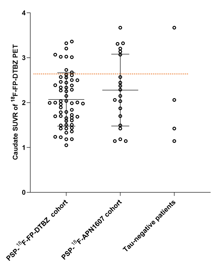
Figure 1
To cite this abstract in AMA style:
C. Dong, J. Ma, S. Liu. Multimodal imaging Integrating 18F-APN-1607 and 18F-FP-DTBZ PET in Progressive Supranuclear Palsy [abstract]. Mov Disord. 2025; 40 (suppl 1). https://www.mdsabstracts.org/abstract/multimodal-imaging-integrating-18f-apn-1607-and-18f-fp-dtbz-pet-in-progressive-supranuclear-palsy/. Accessed July 10, 2026.« Back to 2025 International Congress
MDS Abstracts - https://www.mdsabstracts.org/abstract/multimodal-imaging-integrating-18f-apn-1607-and-18f-fp-dtbz-pet-in-progressive-supranuclear-palsy/