Objective: This study aims to develop a human iPSC-derived oligodendrocyte (iPSC-OL) model to investigate cell-specific disease mechanisms in MSA. Specifically, we seek to:
a)Generate MSA patient-derived iPSC-OLs and characterize their differentiation.
b)Examine the internalization and effects of pathological α-syn fibrils in oligodendrocytes.
c)Establish a platform for transcriptomic profiling to identify disease-related pathways.
Background: MSA is a rare and fatal neurodegenerative disorder characterized by either a parkinsonian (MSA-P) or cerebellar (MSA-C) phenotype, alongside autonomic failure. The etiology remains unknown, and treatments are only symptomatic. One of the key pathological hallmarks of MSA is the prion-like aggregation of α-synuclein (α-syn) in oligodendrocytes (OLs), forming glial cytoplasmic inclusions. While transcriptomic alterations have been identified in postmortem MSA brain tissue, their role in α-syn aggregation and disease progression remains unclear. Developing human cellular models is essential to investigate disease mechanisms.
Method: Fibroblasts from 4 pathologically confirmed MSA cases (CMSAR collection-Table 1) were reprogrammed into iPSCs and were differentiated into OLs along with 2 iPSC control lines using the Douvaras and Fossatti 2015 protocol (Fig 1). Cultures included progenitor and mature OLs, along with neurons and astrocytes from the same iPSC lines. Pathological and physiological α-syn fibrils were extracted and amplified using PMCA from postmortem MSA brains and seeded onto the iPSC-OL cultures. Single-cell capture and indexing of cells was performed using BD Rhapsody SCAnalysis System, followed by single-cell RNA NGS sequencing (scRNA-seq) for transcriptomic profiling.
Results: We successfully generated iPSC-OLs from MSA affected fibroblasts. Confocal microscopy confirmed efficient internalization of pathogenic MSA-brain derived α-syn fibrils within one week, with fibrils predominantly localizing in the perinuclear cytoplasm (see Fig 2). After quality filtering, 3,000–7,000 high-quality cells were identified in the scRNA-seq data, and preliminary analyses show that ~25% of cells expressed oligodendrocyte markers, further validating the differentiation protocol.
Conclusion: This human iPSC-OL model provides a scalable platform for studying MSA pathogenesis. It sets the foundation for future omics analyses to identify disease-specific pathways and potential therapeutic targets.
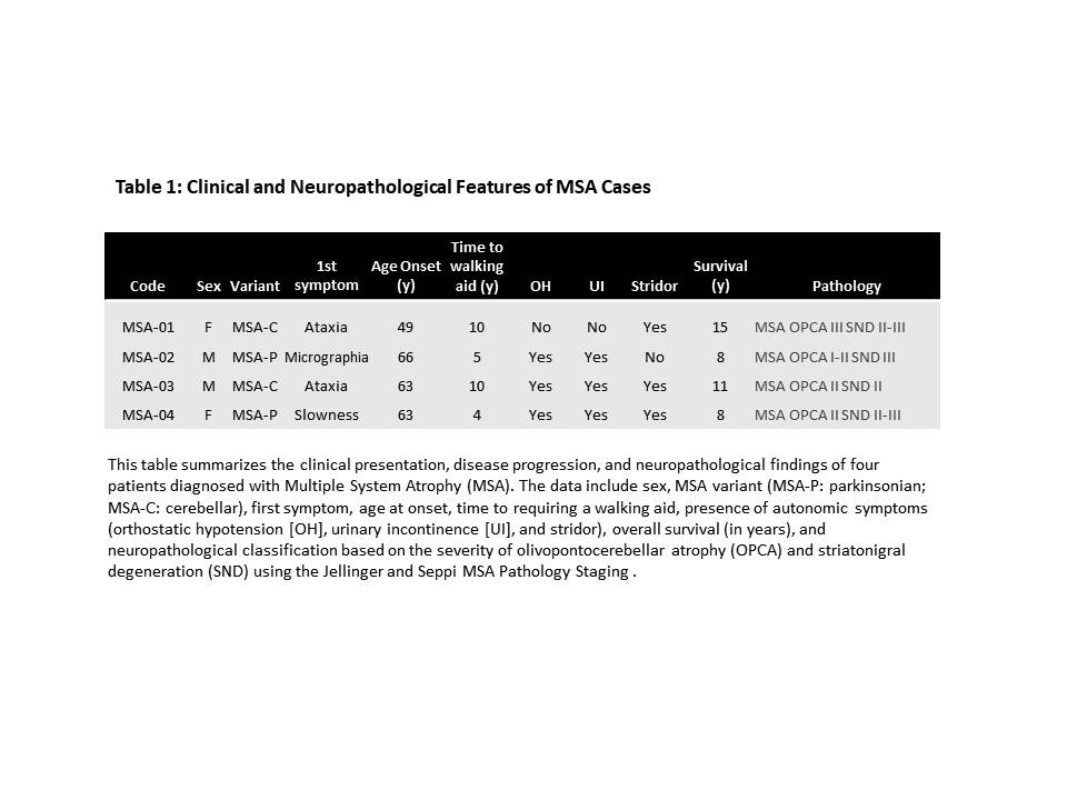
Table 1
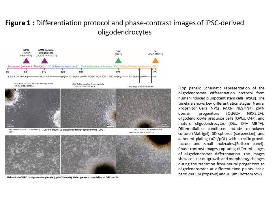
Figure 1
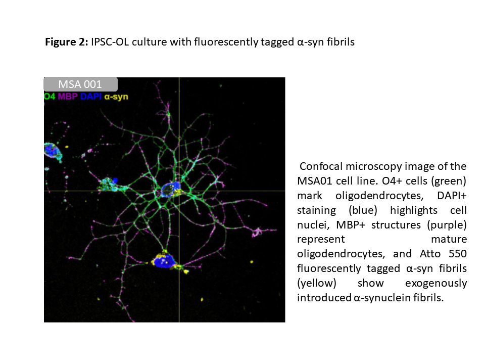
Figure 2
To cite this abstract in AMA style:
A. Perez-Soriano, M. Alemany-Ribes, B. Crespo, F. Kohler, B. Fuste, M. Perez-Soriano, D. Cohen, R. Fernandez-Santiago, M. Ezquerra, MJ. Martí, V. Baekelandt, W. Peelaerts, R. Melki, L. Batlle-Morera, . . Developing a novel Disease-Specific iPSC Model for Studying MSA Pathogenesis [abstract]. Mov Disord. 2025; 40 (suppl 1). https://www.mdsabstracts.org/abstract/developing-a-novel-disease-specific-ipsc-model-for-studying-msa-pathogenesis/. Accessed July 7, 2026.« Back to 2025 International Congress
MDS Abstracts - https://www.mdsabstracts.org/abstract/developing-a-novel-disease-specific-ipsc-model-for-studying-msa-pathogenesis/